

Scanning laser polarimetry with variable and enhanced corneal compensation in normal and glaucomatous eyes
- PMID: 17157800
- PMCID: PMC1832116
- DOI: 10.1016/j.ajo.2006.09.049
Scanning laser polarimetry with variable and enhanced corneal compensation in normal and glaucomatous eyes
Abstract
Purpose: To investigate whether correction for atypical birefringence pattern (ABP) using scanning laser polarimetry with enhanced corneal compensation (SLP-ECC) reduces the severity of ABP compared with variable corneal compensation (SLP-VCC) and improves the correlation with visual function.
Design: Prospective observational study.
Methods: Normal and glaucomatous eyes enrolled from four clinical sites underwent complete examination, automated perimetry, SLP-ECC, and SLP-VCC. Eyes were characterized in three groups based upon the typical scan score (TSS): normal birefringence pattern (NBP) was defined as TSS > or = 80, mild ABP as TSS 61 to 79, and moderate-severe ABP as TSS < or = 60. For each of four SLP parameters, the area under the ROC curve (AUROC) was calculated to compare the ability of SLP-ECC and SLP-VCC to discriminate between normal and glaucomatous eyes.
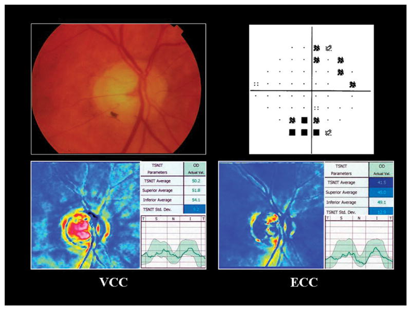
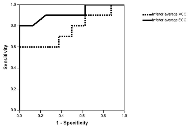
Results: Eighty-four normal volunteers and 45 glaucoma patients were enrolled. Mean TSS was significantly (P < .001) greater using SLP-ECC (98.0 +/- 6.6) compared with SLP-VCC (83.4 +/- 22.5). The frequency of moderate-severe ABP images was significantly (P < .001, McNemar test) higher using SLP-VCC (18 of 129, 14%) compared with SLP-ECC (one of 129, 0.8%). Two SLP-ECC parameters (temporal superior nasal inferior temporal [TSNIT] average and inferior average) had significantly (P = .01, P < .001) higher correlation (r = .45, r = .50, respectively) with MD compared with SLP-VCC (r = .34, r = .37). Among eyes with moderate-severe ABP (n = 18), inferior average obtained using SLP-ECC had significantly (P = .03) greater AUROC (0.91 +/- 0.07) compared with SLP-VCC (0.78 +/- 0.11).
Conclusions: SLP-ECC significantly reduces the frequency and severity of ABP compared with SLP-VCC and improves the correlation between RNFL measures and visual function.
Figures
Comment in
-
Is scanning laser polarimetry ready for clinical practice?Am J Ophthalmol. 2007 Apr;143(4):674-6. doi: 10.1016/j.ajo.2006.12.023. Am J Ophthalmol. 2007. PMID: 17386273 No abstract available.
References
-
- Greenfield DS, Huang X-R, Knighton RW. Effect of corneal polarization axis on assessment of retinal nerve fiber layer thickness by scanning laser polarimetry. Am J Ophthalmol. 2000;129:715–22. - PubMed
-
- Choplin NT, Zhou Q, Knighton RW. Effect of individualized compensation for anterior segment birefringence on retinal nerve fiber layer assessments as determined by scanning laser polarimetry. Ophthalmology. 2003;110:719–25. - PubMed
-
- Huang XR, Knighton RW. Microtubules contribute to the birefringence of the retinal nerve fiber layer. Invest Ophthalmol Vis Sci. 2005;46:4588–93. - PubMed
-
- Huang XR, Knighton RW. Theoretical model of the polarization properties of the retinal nerve fiber layer in reflection. Appl Opt. 2003;42:5726–36. - PubMed
-
- Huang XR, Knighton RW. Diattenuation and polarization preservation of retinal nerve fiber layer reflectance. Appl Opt. 2003;42:5737–43. - PubMed
Publication types
MeSH terms
Grants and funding
LinkOut - more resources
Full Text Sources
Medical
