. 2007 Mar 15;579(Pt 3):703-15.
doi: 10.1113/jphysiol.2006.122432. Epub 2006 Dec 7.

STIM1 regulates Ca2+ entry via arachidonate-regulated Ca2+-selective (ARC) channels without store depletion or translocation to the plasma membrane

Affiliations

STIM1 regulates Ca2+ entry via arachidonate-regulated Ca2+-selective (ARC) channels without store depletion or translocation to the plasma membrane

Olivier Mignen et al. J Physiol. .

Abstract

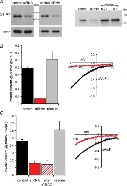
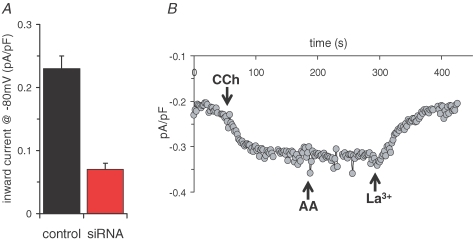
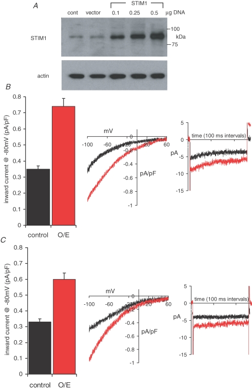
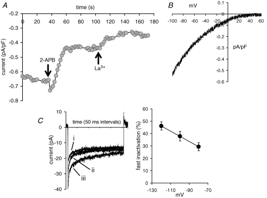
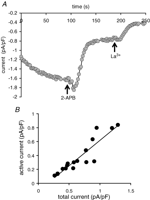
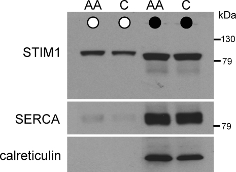
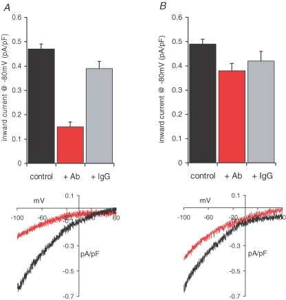
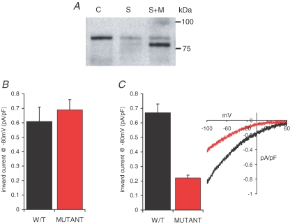
Recent studies have indicated a critical role for STIM (stromal interacting molecule) proteins in the regulation of the store-operated mode of receptor-activated Ca2+ entry. Current models emphasize the role of STIM located in the endoplasmic reticulum membrane, where a Ca2+-binding EF-hand domain within the N-terminal of the protein lies within the lumen and is thought to represent the sensor for the depletion of intracellular Ca2+ stores. Dissociation of Ca2+ from this domain induces the aggregation of STIM to regions of the ER immediately adjacent to the plasma membrane where it acts to regulate the activity of store-operated Ca2+ channels. However, the possible effects of STIM on other modes of receptor-activated Ca2+ entry have not been examined. Here we show that STIM1 also regulates the arachidonic-acid-regulated Ca2+-selective (ARC) channels - receptor-activated Ca2+ entry channels whose activation is entirely independent of store depletion. Regulation of the ARC channels by STIM1 does not involve dissociation of Ca2+ from the EF-hand, or any translocation of STIM1. Instead, a critical role of STIM1 resident in the plasma membrane is indicated. Thus, exposure of intact cells to an antibody targeting the extracellular N-terminal domain of STIM1 inhibits ARC channel activity without significantly affecting the store-operated channels. A similar specific inhibition of the ARC channels is seen in cells expressing a STIM1 construct in which the N-linked glycosylation sites essential for the constitutive cell surface expression of STIM1, were mutated. We conclude that, in contrast to store-operated channels, regulation of ARC channels by STIM1 depends exclusively on the pool of STIM1 constitutively residing in the plasma membrane. These data demonstrate that STIM1 is a more universal regulator of Ca2+ entry pathways than previously thought, and appears to have multiple modes of action.


Figures

Figure 1
Figure 1
siRNA directed against STIM1 inhibits both CRAC channel activity and ARC channel activity in the same cells A, left, representative western blots showing the specific reduction in stromal interacting molecule 1 (STIM1) protein levels in siRNA-expressing cells. Gels were stripped and reprobed with β-actin as a loading control. Right, expression of an siRNA-resistant mutated STIM1 construct (rescue) restores STIM1 protein levels in siRNA-transfected cells. B, the effects of siRNA expression on Ca2+-release-activated Ca2+ (CRAC) channel activity. Mean ±s.e.m. inward current density at −80 mV, and representative current–voltage relationship in cells expressing control siRNA (black, n = 6) are compared with those in cells expressing the siRNA targeted to STIM1 (red, n = 13). Also shown is the effect on mean CRAC currents at −80 mV of expressing an siRNA-resistant construct in the STIM1 siRNA transfected cells (grey, n = 8). CRAC channel currents were activated by use of a Ca2+-free pipette solution containing adenophostin A (2 μm). C, the effects of siRNA expression on arachidonic-acid- regulated Ca2+-selective (ARC) channel activity. Details as in B (control siRNA, n = 6; STIM1 siRNA, n = 11; STIM1 siRNA-resistant, n = 6). Also shown (hatched, n = 6) is the effect on the mean inward ARC channel current density at −80 mV in the same individual siRNA-expressing cells in which reduced CRAC channel currents had been recorded. ARC channel currents were activated by bath addition of arachidonic acid (8 μm).
Figure 2
Figure 2
siRNA directed against STIM1 inhibits agonist-activated ARC channel activity A, the effects of siRNA expression on mean inward currents activated by low (0.5 μm) carbachol concentrations. Mean ±s.e.m. carbachol-activated inward current density at −80 mV in cells expressing control siRNA (black, n = 9) are compared with those in cells expressing the siRNA targeted to STIM1 (red, n = 12). B, representative trace showing the activation of inward current at −80 mV following addition of 0.5 μm carbachol (CCh) to a siRNA-expressing cell. Subsequent addition of arachidonic acid (8 μm) failed to further increase the carbachol-activated current, which was fully blocked by La3+ (100 μm).
Figure 3
Figure 3
Overexpression of STIM1 increases both CRAC channel activity and ARC channel activity A, representative western blot showing STIM1 protein levels in cells transfected with increasing concentrations of a STIM1 construct. Gels were stripped and reprobed with β-actin as a loading control. B, the effects of STIM1 overexpression (0.5 μg DNA) on CRAC channel activity, shown as the mean ±s.e.m. inward current density at −80 mV, in cells transfected with a control vector (black; n = 19) compared with those in cells transfected with the STIM1 construct (red, n = 23). Also shown are representative traces showing the current–voltage relationship and fast inactivation at −80 mV in the CRAC channel currents in control cells (black) and cells overexpressing STIM1 (red). C, the effects of STIM1 overexpression on ARC channel activity. Details as in B (control vector, n = 13; STIM1 overexpression, n = 32). Note the characteristic absence of fast inactivation in the ARC channel currents.
Figure 4
Figure 4
Expression of an EF-hand mutant STIM1 induces the constitutive activation of a current that exclusively reflects CRAC channel activity A, representative trace from a cell expressing the EF-hand mutant STIM1 showing the constitutive activity of an inward current at −80 mV that is partially inhibited by 2-APB (100 μm) and fully inhibited by La3+ (100 μm). The cell was patched with the normal pipette solution (free Ca2+ concentration, 100 nm; see Methods). B, representative trace of the current–voltage relationship of the constitutively active La3+-sensitive current in a cell expressing the EF-hand mutant STIM1. C, the presence of fast inactivation, and its voltage dependence, in the constitutively active La3+-sensitive current in a cell expressing the EF-hand mutant STIM1. Shown are (left) representative recordings showing fast inactivation in the constitutively active current from an individual cell expressing the EF-hand mutant STIM1 recorded during brief (250 ms) pulses to −80 mV (i), −100 mV (ii), and −120 mV (iii), and (right) a plot of the percentage fast inactivation (mean ±s.e.m.) seen in the constitutively active La3+-sensitive current at different voltages in cells expressing the EF-hand mutant STIM1.
Figure 5
Figure 5
Relative contribution of the constitutively active CRAC channel current to total CRAC channel currents in cells expressing the EF-hand mutant STIM1 A, representative trace showing the full activation of CRAC channel currents at −80 mV, and the subsequent effects of 2-APB (100 μm) and La3+ (100 μm) in a cell expressing the EF-hand mutant STIM1. CRAC channels were maximally activated by use of a Ca2+-free pipette solution containing adenophostin (2 μm). B, relationship between the constitutively active current, and the total CRAC channel current measured at −80 mV, for individual cells expressing the EF-hand mutant STIM1.
Figure 6
Figure 6
STIM1 is expressed on the cell surface in the m3-HEK cells Representative western blot showing STIM1 in the biotinylated fraction (^), and whole cell lysate (•) in intact control cells (C) and in cells exposed to 8 μm arachidonic acid (AA). Lysate samples were diluted 4.5-fold compared with the biotinylated samples. The gel was stripped and reprobed with antibodies to SERCA2, and again to calreticulin, as controls for contamination with the endoplasmic reticulum.
Figure 7
Figure 7
Antibodies targeted to the exposed extracellular N-terminal of plasma membrane STIM1 selectively inhibit ARC channel activity A, mean (±s.e.m.) inward current density at −80 mV, and representative current–voltage relationship in azide-control cells (black, n = 11) are compared with those in cells incubated with the N-terminal STIM1 antibody (5 μg ml−1 for 30–40 min at room temperature, red, n = 10). Also shown is the effect of an identical incubation with control IgG2a antibodies (grey, n = 7). B, the effects of the STIM1 antibody on CRAC channel activity, and a representative current–voltage relationship; details as in A; n = 11, 14 and 5, for azide-control cells, STIM1-antibody-treated cells, and control IgG2a-antibody-treated cells, respectively.
Figure 8
Figure 8
Preventing constitutive STIM1 expression on the cell surface selectively inhibits ARC channel activity A, representative western blot showing the expression of the N-glycosylation mutant STIM1 protein levels in siRNA-transfected cells. Shown are STIM1 protein in control cells (C), in cells transfected with the STIM1 siRNA (S), and in siRNA-transfected cells expressing the siRNA-resistant N-glycosylation mutant STIM1 (S + M). Note that the glycosylation mutant runs at a lightly lower molecular mass, and that the siRNA-reduced endogenous STIM1 levels can be seen as a faint band in the same lane running at the normal molecular weight. B, the effects of the N-glycosylation mutant STIM1 on CRAC channel activity. Mean ±s.e.m. inward current density at −80 mV in siRNA-transfected cells expressing the siRNA-resistant wild-type STIM1 (black, n = 8) are compared with those in siRNA-transfected cells expressing the siRNA-resistant N-glycosylation mutant STIM1 (red, n = 7). C, the effects of the N-glycosylation mutant STIM1 on ARC channel activity. Mean ±s.e.m. inward current density at −80 mV (n = 6), and representative current–voltage relationship in siRNA-transfected cells expressing the siRNA-resistant wild-type STIM1 (black) are compared with those in siRNA-transfected cells expressing the siRNA-resistant N-glycosylation mutant STIM1 (red, n = 11).

References

    1. Baba Y, Hayashi K, Fujii Y, Mizushima A, Watarai H, Wakamori M, Numaga T, Mori Y, Iino M, Hikida M, Kursaki T. Coupling of STIM1 to store-operated Ca2+ entry through its constitutive and inducible movement in the endoplasmic reticulum. Proc Natl Acad Sci U S A. 2006;103:16704–16709. - PMC - PubMed
    1. Berridge MJ. Inositol trisphosphate and calcium signalling. Nature. 1993;361:315–325. - PubMed
    1. Bootman MD, Berridge MJ, Roderick HL. Calcium signalling: more messengers, more channels, more complexity. Curr Biol. 2002;12:R563–R565. - PubMed
    1. Hermosura MC, Monteilh-Zoller MK, Scharenberg AM, Penner R, Fleig A. Dissociation of the store-operated calcium current ICRAC and the Mg-nucleotide-regulated metal ion current MagNuM. J Physiol. 2002;539:445–458. - PMC - PubMed
    1. Hoth M, Penner R. Depletion of intracellular calcium stores activates a calcium current in mast cells. Nature. 1992;355:353–356. - PubMed

Publication types

MeSH terms

LinkOut - more resources