Review
. 2006 Dec;70(4):910-38.
doi: 10.1128/MMBR.00020-06.

Stimulus perception in bacterial signal-transducing histidine kinases

Affiliations
Review

Stimulus perception in bacterial signal-transducing histidine kinases

Thorsten Mascher et al. Microbiol Mol Biol Rev. 2006 Dec.

Abstract

Two-component signal-transducing systems are ubiquitously distributed communication interfaces in bacteria. They consist of a histidine kinase that senses a specific environmental stimulus and a cognate response regulator that mediates the cellular response, mostly through differential expression of target genes. Histidine kinases are typically transmembrane proteins harboring at least two domains: an input (or sensor) domain and a cytoplasmic transmitter (or kinase) domain. They can be identified and classified by virtue of their conserved cytoplasmic kinase domains. In contrast, the sensor domains are highly variable, reflecting the plethora of different signals and modes of sensing. In order to gain insight into the mechanisms of stimulus perception by bacterial histidine kinases, we here survey sensor domain architecture and topology within the bacterial membrane, functional aspects related to this topology, and sequence and phylogenetic conservation. Based on these criteria, three groups of histidine kinases can be differentiated. (i) Periplasmic-sensing histidine kinases detect their stimuli (often small solutes) through an extracellular input domain. (ii) Histidine kinases with sensing mechanisms linked to the transmembrane regions detect stimuli (usually membrane-associated stimuli, such as ionic strength, osmolarity, turgor, or functional state of the cell envelope) via their membrane-spanning segments and sometimes via additional short extracellular loops. (iii) Cytoplasmic-sensing histidine kinases (either membrane anchored or soluble) detect cellular or diffusible signals reporting the metabolic or developmental state of the cell. This review provides an overview of mechanisms of stimulus perception for members of all three groups of bacterial signal-transducing histidine kinases.


Figures

FIG. 1.
FIG. 1.
Schematic representation of the three different mechanisms of stimulus perception. (A) Periplasmic-sensing HKs. (B) HKs with sensing mechanisms linked to the transmembrane regions (stimulus perception can occur either with the membrane-spanning helices alone or by combination of the transmembrane regions and short extracellular loops). (C) Cytoplasmic-sensing HKs (either soluble or membrane-anchored proteins). The stimulus is represented by a red arrow or red star. The parts of the proteins involved in stimulus perception are highlighted in color.
FIG. 2.
FIG. 2.
Features, domains, and boxes of histidine protein kinases. The protein is symbolized by the gray line. The major domains are indicated by boxes, and their names are given above or below the line. Conserved boxes or amino acid residues are given below the line in one-letter code, according to the standard nomenclature (87, 280). The drawing is not to scale. See the text and Table 1 for details.
FIG. 3.
FIG. 3.
Domain architecture of periplasmic-sensing histidine kinases. The figure is based on the graphical output of the SMART web interface at http://smart.embl-heidelberg.de (229), with modifications. The scale bar is in amino acids. Blue vertical bars represent putative transmembrane helices. Sizes and positions of conserved domains are indicated by the labeled symbols. Note that the transmitter domains are simplified, and as a default, only the HisKA and HATPase_c domains are shown. Additional cytoplasmic domains are possible and widespread but were ignored in all but obligatory cases (i.e., PAS domain for CitA-like HKs and HAMP domain for NarXQ-like HKs). A diagonal bar at the C terminus of the transmitter domain indicates the possible occurrence of hybrid kinases in that subgroup of sensor kinases. The periplasmic PAS domain of CitA/DcuS (in parentheses) is conserved by three-dimensional structure only and not by sequence. It is therefore not detectable by sequence analysis. VanS/PrmB-like proteins are described in the “Intramembrane-Sensing HKs: Cell Envelope Stress Sensors with Two TMR (LiaS/BceS-Like HKs)” section. See the text for details.
FIG. 4.
FIG. 4.
Structure of the periplasmic input domain of PhoQ and model for the sensing mechanism of cations and antimicrobial peptides. (A) The crystal structure and charge profile of the surface facing the outer side of the cytoplasmic membrane are shown on the left. The residues important for coordinating the divalent metal ions are shown. The crystal structure of the dimeric PhoQ sensor domain (upper panel) forms a flat surface that comes in close contact to the membrane. The bottom part of this domain contains a highly negatively charged surface that participates in metal binding (lower panel, view from the membrane). Red represents negatively charged residues. NT, N terminus; CT, C terminus. (B) Working model for the competitive binding of Mg2+ and cationic antimicrobial peptides to PhoQ. Divalent cations, such as Ca2+ or Mg2+ (shown as green balls), bind to the acidic surface (red) and repress PhoQ activity by locking the PhoQ sensor domain in an inactive conformation (top panel). Cationic antimicrobial peptides interact with membrane phospholipids, thereby coming in close contact with the Ca2+ and Mg2+ binding sites of PhoQ. They compete with and displace divalent cations from PhoQ (middle panel). This provokes a conformational change of the input domain, which leads to autophosphorylation of the transmitter domain and thereby activation of PhoQ (lower panel). See the text for details. (Reprinted from reference with permission from Elsevier.)
FIG. 5.
FIG. 5.
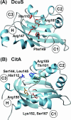
Structures of the periplasmic sensing domains of DcuS (A) and CitA (B). The structures for the periplasmic domains of CitA and DcuS are derived from http://www.rcsb.org. The residues required for C4-dicarboxylate sensing by DcuS (144, 201) and direct binding of citrate to CitA (75, 219) are shown. The corresponding sites are highlighted in the structure.
FIG. 6.
FIG. 6.
Domain architecture of histidine kinases with sensing mechanisms linked to the transmembrane regions. The figure is based on the graphical output of the SMART web interface at http://smart.embl-heidelberg.de (229), with modifications. The scale bar is in amino acids. Blue vertical bars represent putative transmembrane helices. Sizes and positions of conserved domains are indicated by the labeled symbols. Note that the transmitter domains are simplified, and as a default, only HisKA and HATPase_c are shown. See the text for details.
FIG. 7.
FIG. 7.
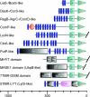
Domain architecture of some examples of cytoplasmic-sensing histidine kinases. The figure is based on the graphical output of the SMART web interface at http://smart.embl-heidelberg.de (229), with modifications. The scale bar is in amino acids. Blue vertical bars represent putative transmembrane helices. Sizes and positions of conserved domains are indicated by the labeled symbols. See the text for details.
FIG. 8.
FIG. 8.
Model for the redox regulation of the ArcB sensor kinase of E. coli through inactivation by oxygen. Upon a shift from anaerobic to aerobic growth conditions, the respiratory quinone (ubiquinone [Q]) becomes oxidized by oxygen via the respiratory chain. The oxidized quinones oxidize Cys180 of ArcB, resulting in an intermolecular Cys180/Cys180 disulfide bridge of the ArcB dimer. The resulting conformational changes lead to a reduced kinase activity of ArcB. Under fully oxic conditions, when the quinones are maximally oxidized by the respiratory chain, the quinones oxidize Cys241 of the ArcB linker domain. The additional conformational changes completely silence ArcB kinase activity. (Reprinted from reference with permission of the publisher. Copyright 2004 National Academy of Sciences, U.S.A.).

References

    1. Abada, E. M., A. Balzer, A. Jager, and G. Klug. 2002. Bacteriochlorophyll-dependent expression of genes for pigment-binding proteins in Rhodobacter capsulatus involves the RegB/RegA two-component system. Mol. Genet. Genomics 267:202-209. - PubMed
    1. Aguilar, P. S., J. E. Cronan, Jr., and D. de Mendoza. 1998. A Bacillus subtilis gene induced by cold shock encodes a membrane phospholipid desaturase. J. Bacteriol. 180:2194-2200. - PMC - PubMed
    1. Aguilar, P. S., A. M. Hernandez-Arriaga, L. E. Cybulski, A. C. Erazo, and D. de Mendoza. 2001. Molecular basis of thermosensing: a two-component signal transduction thermometer in Bacillus subtilis. EMBO J. 20:1681-1691. - PMC - PubMed
    1. Aguilar, P. S., P. Lopez, and D. de Mendoza. 1999. Transcriptional control of the low-temperature-inducible des gene, encoding the Δ5 desaturase of Bacillus subtilis. J. Bacteriol. 181:7028-7033. - PMC - PubMed
    1. Anantharaman, V., and L. Aravind. 2003. Application of comparative genomics in the identification and analysis of novel families of membrane-associated receptors in bacteria. BMC Genomics 4:34. - PMC - PubMed

Publication types

MeSH terms

LinkOut - more resources