. 2006 Dec 1;20(23):3238-43.
doi: 10.1101/gad.1496606.

MDC1 accelerates nonhomologous end-joining of dysfunctional telomeres

Affiliations

MDC1 accelerates nonhomologous end-joining of dysfunctional telomeres

Nadya Dimitrova et al. Genes Dev. .

Abstract

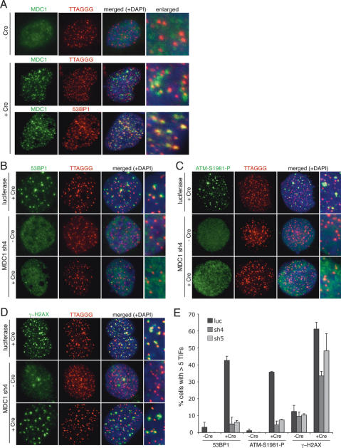
Here we document the role of MDC1 (mediator of DNA damage checkpoint 1) in the detection and repair of human and mouse telomeres rendered dysfunctional through inhibition of TRF2. Consistent with its role in promoting DNA damage foci, MDC1 knockdown affected the formation of telomere dysfunction-induced foci (TIFs), diminishing the accumulation of phosphorylated ATM, 53BP1, Nbs1, and to a lesser extent, gamma-H2AX. In addition to this effect on TIFs, the rate of nonhomologous end-joining (NHEJ) of dysfunctional telomeres was significantly decreased when MDC1 itself or its recruitment to chromatin was inhibited. MDC1 appeared to promote a step in the NHEJ pathway after the removal of the 3' telomeric overhang. The acceleration of NHEJ was unlikely to be due to increased presence of 53BP1 and Mre11 in TIFs, since knockdown of neither factor affected telomere fusions. Furthermore, relevant cell cycle effectors (Chk2, p53, and p21) of the ATM kinase pathway were unaffected and there was no change in the rate of cell cycle progression. We propose that the binding of MDC1 to gamma-H2AX directly affects NHEJ in a manner that is independent of the ATM-dependent cell cycle arrest pathway.


Figures

Figure 1.
Figure 1.
MDC1 localizes at dysfunctional telomeres and promotes the formation of TIFs. (A) MDC1 recruitment to chromosome ends after deletion of a conditional TRF2 allele in TRF2F/− lig4−/− p53−/− MEFs. Cells untreated or treated with Cre were fixed 72 h post-infection and processed for IF-FISH (MDC1 [green] costained with telomeric TTAGGG-specific FISH probe [red]) or IF (MDC1 [green] costained with 53BP1 [red]). (B–D) Effect of MDC1 knockdown on 53BP1 (B), ATM-S1981-P (C), and γ-H2AX (D) TIF formation in mouse cells. TRF2F/− lig4−/− p53−/− MEFs expressing control luciferase or mouse MDC1-specific shRNA, sh4, were fixed 72 h after mock or Cre infection and processed for IF-FISH (53BP1, ATM-S1981-P, and γ-H2AX [green]; TTAGGG probe [red]; and DNA counterstained with DAPI [blue]). (E) Bar graph of the frequency of TIF formation. At least 100 cells were scored for five or more telomeric 53BP1, ATM-S1981-P, and γ-H2AX foci in two independent experiments.
Figure 2.
Figure 2.
MDC1 stimulates NHEJ of dysfunctional telomeres. (A) Metaphase spreads from TRF2F/− p53−/− and TRF2F/+ p53−/− MEFs expressing control luciferase or mouse MDC1-specific shRNA, sh4, 60 h after Cre infection. Telomeric signals were detected with FITC-OO-(AATCCC)3 oligonucleotide and are false-colored in green; DNA (DAPI) is false-colored in red. (B) Quantification of telomere fusions in TRF2F/− p53−/− MEFs, expressing the indicated shRNA, mock- or Cre-infected for 60–72 h. Telomere fusions were counted as events per chromosome in metaphase spread as shown in A. Bar graph represents fusion frequency relative to control shRNA-treated cells in three independent experiments. (C) Telomeric DNA analysis. (Left) In-gel assay detecting 3′ overhang of TRF2F/− p53−/− MEFs expressing control luciferase or MDC1 shRNAs, sh4 and sh5. Cells were harvested 72 h post-infection with Cre and processed by in-gel hybridization to a (CCCTAA)4 probe to detect ssTTAGGG repeats (native). (Right) The DNA was denatured in situ and rehybridized to the same probe to detect the total TTAGGG signal (denatured). Overhang signals were quantified with ImageQuant software and normalized to the total TTAGGG signal in the same lane. The numbers below the gel represent the percentage of normalized overhang signal compared with the normalized overhang signal for the same cells not treated with Cre. (D) Quantification of telomeric overhang 72, 96, and 120 h post-infection with Cre in two independent experiments. The overhang signal at different time points after Cre infection is represented as a percentage of the overhang signal in the absence of Cre for the same cell line.
Figure 3.
Figure 3.
Recruitment of MDC1 to chromatin is required for efficient NHEJ. (A) Immunoblot detection of H2AX in HeLa cells expressing control or H2AX-specific shRNA, sh3; nonspecific band from the same gel used as loading control. (B) Effect of H2AX knockdown on MDC1 TIF formation. HeLa cells expressing control luciferase or H2AX-specific shRNA were fixed 48 h post-infection with control (β-gal) or TRF2-DN adenovirus and processed for IF (γ-H2AX [green]; MDC1 [red]; and DNA counterstained with DAPI [blue]). (C) At least 100 cells were scored for five or more telomeric MDC1 foci in three independent experiments. (D) Metaphase spreads from HeLa cells expressing control luciferase or H2AX-specific shRNA 48 h post-infection with TRF2-DN or control (β-gal) adenovirus. Telomeric signals were detected with FITC-OO-(AATCCC)3 oligonucleotide and are false-colored in green; DNA (DAPI) is false-colored in red. (E) Quantification of telomere fusions in C counted as events per chromosome end in three independent experiments. All the results were confirmed with a second independent H2AX shRNA.
Figure 4.
Figure 4.
MDC1 knockdown does not affect checkpoint signaling in response to telomere dysfunction. (A) Immunoblot detection of ATM-S1981-P and Chk2-P after telomere deprotection. Whole-cell lysates of TRF2F/− lig4−/− p53−/− MEFs expressing control luciferase or MDC1-specific shRNAs, sh4 and sh5, were analyzed 72 h after mock or Cre infection. TRF2 and Rap1 immunoblots confirm efficient TRF2 deletion. (B) Detection of up-regulation of p53 and p21 in primary cells. Vector or MDC1 shRNA-treated IMR90 cells were harvested for immunoblot analysis 48 h post-infection with TRF2-DN (AdDN), the β-gal adenovirus control (AdβG), or 1 h post-irradiation with 5 Gy (IR). The MDC1 immunoblot confirmed knockdown of MDC1 by two independent shRNAs; TRF2-DN expression is detected with a myc antibody. (C) Microscopic images of IMR90 cells, expressing vector or MDC1 shRNA, sh1, infected with empty vector control or TRF2-DN retrovirus stained for SA-β-galactosidase activity at 12 d after infection. (D) Lack of effect of MDC1 shRNA on proliferation of MEFs. Growth curve of TRF2F/− p53−/− MEFs after Cre infection. Inset table represents percent of BrdU-positive cells 72 h after mock or Cre infection.

References

    1. Bekker-Jensen, S., Lukas, C., Melander, F., Bartek, J., Lukas, J. Dynamic assembly and sustained retention of 53BP1 at the sites of DNA damage are controlled by Mdc1/NFBD1. J. Cell Biol. 2005;170:201–211. - PMC - PubMed
    1. Bekker-Jensen, S., Lukas, C., Kitagawa, R., Melander, F., Kastan, M.B., Bartek, J., Lukas, J. Spatial organization of the mammalian genome surveillance machinery in response to DNA strand breaks. J. Cell Biol. 2006;173:195–206. - PMC - PubMed
    1. Celli, G., de Lange, T. DNA processing not required for ATM-mediated telomere damage response after TRF2 deletion. Nat. Cell Biol. 2005;7:712–718. - PubMed
    1. Celli, G.B., Lazzerini Denchi, E., de Lange, T. Ku70 stimulates fusion of dysfunctional telomeres yet protects chromosome ends from homologous recombination. Nat. Cell Biol. 2006;8:885–890. - PubMed
    1. di d’Adda Fagagna, F., Reaper, P.M., Clay-Farrace, L., Fiegler, H., Carr, P., Von Zglinicki, T., Saretzki, G., Carter, N.P., Jackson, S.P. A DNA damage checkpoint response in telomere-initiated senescence. Nature. 2003;426:194–198. - PubMed

Publication types

MeSH terms