

Substance P is responsible for physiological alterations such as increased chloride ion secretion and glucose malabsorption in cryptosporidiosis
- PMID: 17158891
- PMCID: PMC1828596
- DOI: 10.1128/IAI.01738-05
Substance P is responsible for physiological alterations such as increased chloride ion secretion and glucose malabsorption in cryptosporidiosis
Abstract
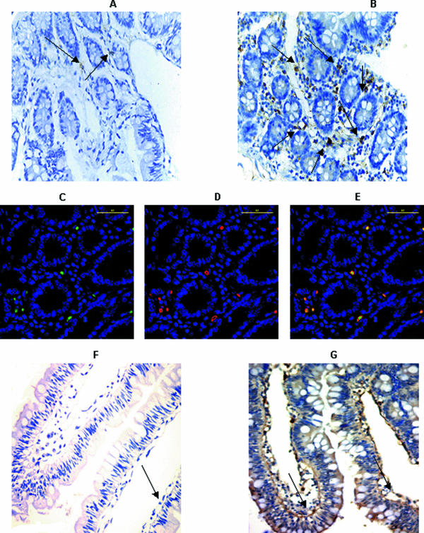
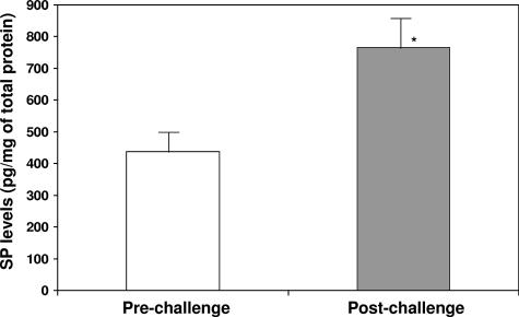
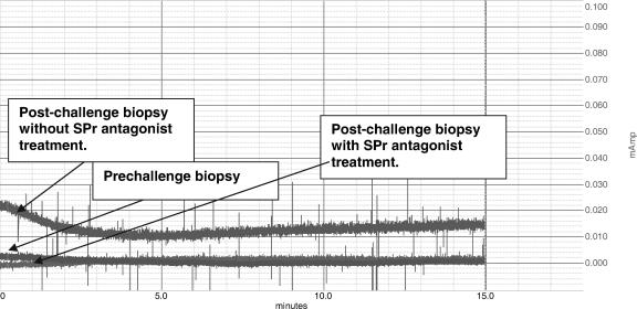
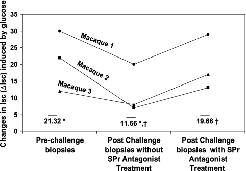
Cryptosporidiosis, caused by the protozoan parasite Cryptosporidium, causes self-limited diarrhea in immunocompetent hosts and severe life-threatening diarrhea in AIDS patients. Highly active antiretroviral therapy has been used to effectively treat cryptosporiosis in some but not all AIDS patients. Therefore, there is an urgent need for innovative drugs to treat this disease. Cryptosporidium infection results in intestinal pathophysiological changes such as glucose malabsorption, increased chloride ion (Cl(-)) secretion, and epithelial barrier disruption, leading to disease pathogenesis. In order to develop tools to combat this opportunistic pathogen, it is vital to understand mediators involved in disease pathogenesis. Substance P (SP), a neuropeptide and pain transmitter, is located in the gastrointestinal tract. SP can cause Cl(-) secretion in human gastrointestinal explants. However, its role in cryptosporidiosis has not been fully studied. Jejunal samples from macaques before and after Cryptosporidium parvum infection were assayed for SP and SP receptor mRNA and protein levels by reverse transcription-PCR and by immunohistochemistry and enzyme-linked immunosorbent assay, respectively. The role of SP in pathophysiological alterations, such as Cl(-) secretion and glucose malabsorption, was studied using tissues derived from macaques infected with C. parvum by the Ussing chamber technique. SP and SP receptor mRNA and protein expression levels were increased in jejunal samples following C. parvum infection and were accompanied by increased basal ion secretion and glucose malabsorption. In vitro treatment of samples obtained from infected macaques with the SP receptor antagonist aprepitant (Emend; Merck, Whitehouse Station, NJ) completely reversed the increase in basal ion secretion and corrected the glucose malabsorption. Our findings raise the possibility of using SP receptor antagonists for the treatment of symptoms associated with cryptosporidiosis.
Figures
References
-
- Abe, Y., N. A. Bui-Thanh, C. M. Ballantyne, and A. R. Burns. 2005. Extra domain A and type III connecting segment of fibronectin in assembly and cleavage. Biochem. Biophys. Res. Commun. 338:1640-1647. - PubMed
-
- Anton, P. A., and F. Shanahan. 1998. Neuroimmunomodulation in inflammatory bowel disease. How far from “bench” to “bedside”? Ann. N. Y. Acad. Sci. 840:723-734. - PubMed
-
- Argyropoulos, S. V., and D. J. Nutt. 2000. Substance P antagonists: novel agents in the treatment of depression. Expert Opin. Investig. Drugs 9:1871-1875. - PubMed
-
- Berczi, I., I. M. Chalmers, E. Nagy, and R. J. Warrington. 1996. The immune effects of neuropeptides. Baillieres Clin. Rheumatol. 10(2):227-257. - PubMed
-
- Bernstein, C. N., M. E. Robert, and V. E. Eysselein. 1993. Rectal substance P concentrations are increased in ulcerative colitis but not in Crohn's disease. Am. J. Gastroenterol. 88:908-913. - PubMed
Publication types
MeSH terms
Substances
Grants and funding
LinkOut - more resources
Full Text Sources
Medical
