. 2006 Dec 15;119(Pt 24):5124-36.
doi: 10.1242/jcs.03292.

Multiple controls regulate nucleostemin partitioning between nucleolus and nucleoplasm

Affiliations

Multiple controls regulate nucleostemin partitioning between nucleolus and nucleoplasm

Lingjun Meng et al. J Cell Sci. .

Abstract

Nucleostemin plays an essential role in maintaining the continuous proliferation of stem cells and cancer cells. The movement of nucleostemin between the nucleolus and the nucleoplasm provides a dynamic way to partition the nucleostemin protein between these two compartments. Here, we show that nucleostemin contains two nucleolus-targeting regions, the basic and the GTP-binding domains, that exhibit a short and a long nucleolar retention time, respectively. In a GTP-unbound state, the nucleolus-targeting activity of nucleostemin is blocked by a mechanism that traps its intermediate domain in the nucleoplasm. A nucleostemin-interacting protein, RSL1D1, was identified that contains a ribosomal L1-domain. RSL1D1 co-resides with nucleostemin in the same subnucleolar compartment, unlike the B23 and fibrillarin, and displays a longer nucleolar residence time than nucleostemin. It interacts with both the basic and the GTP-binding domains of nucleostemin through a non-nucleolus-targeting region. Overexpression of the nucleolus-targeting domain of RSL1D1 alone disperses nucleolar nucleostemin. Loss of RSL1D1 expression reduces the compartmental size and amount of nucleostemin in the nucleolus. Our work reveals that the partitioning of nucleostemin employs complex mechanisms involving both nucleolar and nucleoplasmic components, and provides insight into the post-translational regulation of its activity.


Figures

Figure 1
Figure 1. Nucleostemin (NS) contained two distinct nucleolus-targeting regions with different nucleolar retention time
(A) A schematic diagram of NS protein structure and mutant constructs used to determine the nucleolus-targeting domains of NS. An SV40 nuclear localization signal (NLS, black circle) was engineered in mutants missing the endogenous NLS (black boxes). Numbers indicate the amino acid positions. Abbreviations: B, basic; C, coiled-coil; G, GTP-binding; I, intermediate; A, acidic. (B) The subcellular distributions of mutant proteins in U2OS cells were revealed by a C-terminal green fluorescent protein (GFP) tag, and counterstained with anti-B23 immunofluorescence shown in the right upper quadrants on a 50% scale. Both the B- and the G-domains (nlsG) displayed a nucleolar distribution pattern. A mutation in the conserved GTP-binding residue G256 (nlsG(256)) did not affect the nucleolar localization of the G-domain alone. When the I-domain was present, such a mutation would abolish its nucleolar localization (nlsGI(256)). A cytoplasmic hydrolase protein (Hd3) was tagged with the SV40 NLS to demonstrate that this sequence was not sufficient to confer nucleolar localization. Scale bar: 10 um. (C1) The FRAP (fluorescence recovery after photobleaching) recovery time (X-axis, in seconds) of the B-domain (trace 1), the full-length NS (trace 2), and the GI-domain (trace 3) was determined in Chinese hamster ovary (CHO) cells transiently transfected with the GFP-fusion constructs. The Y-axis represented the percentage of the fluorescence intensity in the bleached area relative to the prebleached intensity. (C2) The FRAP recovery time of the GI-domain (nlsGI) and the A-domain deletion mutant (NSdA) was measured as described in the methods section. Statistical analyses at 5, 10, 20, and 30 seconds were shown in (C3) (mean ± standard error of mean (s.e.m), n=20).
Figure 2
Figure 2. The nucleolar localization of NS was gated by a nucleoplasmic-retaining mechanism independent of its nucleolus-targeting domains
When fused to the N-terminus (A1) or the C-terminus (B1) of B23, the I-domain of NS significantly increased the amount of B23 in the nucleoplasm, compared to the epitope-tagged (A2, B2) or the GFP-tagged proteins (A3, B3) at their respective ends. This nucleoplasmic-retaining activity of the I-domain could also be transferred to three ribosomal proteins, L5, L11, and L23. Unlike the nucleolar distributions of their original proteins (C2, D2, E2), the I-domain fusions of these proteins (C1, D1, E1) were localized almost exclusively in the nucleoplasm. Anti-fibrillarin or anti-B23 immunostainings of the same cells were shown in the bottom panels. Fusion constructs were depicted at the bottom of each panel. Scale bar: 10 um.
Figure 3
Figure 3. NS interacted with a ribosomal L1 domain-containing protein 1, RSL1D1
(A) Protein sequences of mouse RSL1D1 (NM_025546) and two closely related genes, L10A (NM_011287), and L10 (XM_138143), were aligned by the Clustal W (1.81) program. The shaded and underlined areas represented the ribosomal L1 and the coiled-coil domain, respectively. Three putative NLS were marked in bold letters. Consensus keys: fully conserved residues, asterisk; conservation of strong groups, double dots; conservation of weak groups, single dot. Biochemical interaction between NS and RSL1D1 was shown by affinity binding assays using GST fusion of RSL1D1 to pull down HA-tagged NS (B1) or GST fusion of NS to pull down HA-tagged RSL1D1 (B2). (C) The NS-RSL1D1 interaction was confirmed by coimmunoprecipitation in both directions. HEK293 cells were cotransfected with HA-tagged NS and myc-tagged RSL1D1 and immunoprecipitated with anti-myc antibody (1st and 2nd rows, left column), anti-HA antibody (3rd and 4th rows, left column), or mouse IgG (1st to 4th rows, right column). The co-purified proteins (1st and 3rd rows) and the immunoprecipitates (2nd and 4th rows) were detected by immunoblotting with the indicated polyclonal antibodies. (D) Myc-tagged RSL1D1 could be co-purified with endogenous NS in HEK293 cells by α-NS antiserum (Ab1164), but not by preimmune serum (Cntrl) (left panel). Endogenous NS could also be co-purified with myc-tagged RSL1D1 by α-myc antibody, but not by mouse IgG (right panel).
Figure 4
Figure 4. Tissue and subcellular distributions of RSL1D1 overlapped with those of NS
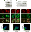
The expression patterns of RSL1D1 and NS in the developing whole embryos from embryonic day 10.5 (E10.5) to E16.5 (A) and in the adult mice (B) were shown by Northern blot analyses. Colocalization of endogenous NS (red, detected by Ab2438) and myc-tagged RSL1D1 (green), NS (red) and B23 (green), and NS (red) and fibrillarin (green, Fib) were shown by double-labeled immunofluorescence and confocal analyses in (C), (D), and (E), respectively. High magnifications of the indicated areas (squares) were shown in (C2, D2, and E2). Colocalization was quantified in (C3, D3, and E3) where all pixels were plotted based on their red (X-axis) and green (Y-axis) fluorescence intensities, and pseudocolored based on the event frequency, with red representing the highest and blue representing the lowest event frequency. Dashed lines delineate the nucleo-cytoplasmic boundaries. Scale bars: 5 um for C1, D1, and E1; 2 um for C2, D2, and E2.
Figure 5
Figure 5. RSL1D1 had a longer nucleolar retention time than NS
(A) Time-sequenced FRAP images of NS and RSL1D1 in the nucleolus. A circle of 1um in diameter within the nucleolus (marked by arrows) was bleached. Of note, low intensity spots in the upper panels (indicated by asterisk) existed before photobleaching. Numbers indicate time (in seconds) after the bleaching event. Scale bar: 1 um. (B) The FRAP recovery curves of RSL1D1 and NS depicted the average of the fluorescence recovery level (Y-axis, n=20) relative to the prebleached intensity (set as 1) over a 31.6-second period following photobleaching (X-axis, in seconds). Y-error bars represented standard deviations (s.d.) and were omitted on the top and bottom side of the RSL1D1 and NS recovery curves for clarity. (C) T-test analyses of the FRAP results were conducted at 5, 10, 20, and 30 seconds after photobleaching (means ± s.e.m, n=20).
Figure 6
Figure 6. RSL1D1 interacted with the B- and G-domains of NS
(A) Schematic diagrams of NS truncated mutants used to determine its RSL1D1-interacting domain(s). (B) Affinity binding assays showed that GST fusions of RSL1D1 were able to pull down all single-domain deletion mutants of NS, suggesting that multiple regions were involved. (C) Using complex deletion mutants, we showed that RSL1D1 was able to bind both the BC- and the GI-domains, but noy the G-, IA-, or GI-domains with a G256V mutation (GI(256)). The RSL1D1-binding domains of NS were further defined to the B-region of the BC-domain (D1) and the GI1-region of the GI-domain (D2). (D2) Double deletion mutants (NSdBG and NSdCI) confirmed the importance of the B- and G-domains, but not the C- and I-domains, in mediating the NS-RSL1D1 interaction.
Figure 7
Figure 7. The nucleolar distribution and the NS-interaction of RSL1D1 were controlled by separate domains
(A) RSL1D1 contained an L1 domain (amino acid 150–254), a coiled-coil domain (C), and three putative NLS (black boxes). Myc-tagged RSL1D1 truncated mutants were generated to map its NS-interacting and nucleolus-targeting regions. (B) Affinity binding assays showed that GST fusions of both the BC- and GI-domains could bind the (317–452) portion of RSL1D1, which did not contain the L1- or C-domain. (C-L) Anti-myc and anti-NS (Ab2438) double-labeled immunofluorescence demonstrated that the 150–316 region of RSL1D1 was localized in the nucleolus (C). The N-terminal 1–149 region was cytoplasmic by itself (D), and became partially nucleolar when tagged with an SV40 NLS (E). The distribution of the C-terminal 317–452 region was diffuse in the nucleus (F), with some cells showing more signals in the nucleolus than in the nucleoplasm (G). Within the 150–316 segment, the L1 domain (150–254) was primarily cytoplasmic by itself (H), but became mostly nucleolar when provided with a SV40 NLS (I, J, nlsL1). The coiled-coil domain (255–316) was diffusely localized in the nucleus (K). The NS signal was diminished or absent from the nucleolus of many cells expressing nlsL1 (J). Scale bar: 10 um.
Figure 8
Figure 8. Overexpression of a nucleolar form of the L1-domain (nlsL1) dispersed NS from the nucleolus
(A1) The intensities of NS signals in the nucleolus were diminished or disappeared in many cells expressing the nlsL1 mutant. (A2) High magnification of the nlsL1-expressing cell showed that its remaining NS signals displayed a reticular pattern of distribution, similar to the NS distribution in wild-type cells. (B) In some cells, NS was scattered around the nlsL1 signals. Overexpression of the nlsL1 mutant did not affect the signal intensities or the distribution patterns of fibrillarin (C) or B23 (D). Scale bars: 10 um for A1; 5 um for A2, B, C, D.
Figure 9
Figure 9. Loss of RSL1D1 expression decreased the compartmental size and protein amount of NS and B23 in the nucleolus
(A1) The knockdown efficiency of the RSL1D1-specific siRNA duplex (siRSL1D1) was examined at the RNA and protein levels. Compared to samples treated with the control siRNA duplex (siNEG), siRSL1D1 was able to reduce the endogenous RSL1D1 mRNA by 73% (top panel), and the exogenously expressed myc-tagged RSL1D1 protein by 43% in HEK293 cells (bottom panel). The siRSL1D1 treatment did not affect the total protein amount of NS (A2). Tub: β-tubulin for Northern blots (NB), and α-tubulin for Western blots. The effect of a partial loss of RSL1D1 expression on the nucleolar distribution of NS was measured in U2OS cells double-labeled with anti-NS (Ab2438) and anti-B23 immunofluorescence. Quantitative analyses showed that a partial knockdown of RSL1D1expression decreased the total nucleolar area (No) occupied by NS (B1) (p < 0.001, n=130). A similar effect was seen in the B23-containing regions (B2). The Y-axis represents the percentage of cells at or below the size indicated on the X-axis. The X-axis represents the nucleoluar area in units of 100 pixels (= 0.89 um2). (C) Immunofluorescence images representative of the average of each group were shown. Dashed lines delineate the nucleo-cytoplasmic boundaries. Scale bars: 5 um.
Figure 10
Figure 10. Schematic diagrams of the mechanism controlling NS distribution between the nucleolar and nucleoplasmic compartments, and the effects of RSL1D1 knockdown and nlsL1 overexpression
Our work demonstrates that NS in the GTP-unbound state is blocked from entering the nucleolus by a nucleoplasmic-retaining mechanism that acts on the I-domain. GTP binding releases this lock and allows NS to move into the nucleolus. NS interacts with nucleolar protein RSL1D1 through the nucleolus-targeting B- and G-domains. When not bound by GTP, the GI-domain fails to interact with RSL1D1, suggesting a link between the nucleolar exit of NS and GTP hydrolysis. RSL1D1 co-resides with NS in the same subnucleolar domains surrounding fibrillarin. A partial knockdown of RSL1D1 expression reduces the compartmental size and, to a lesser extent, the protein amount of NS in the nucleolus, supporting with idea that RSL1D1 provides the nucleolar binding site for NS. Overexpression of nlsL1 disperses NS signals from the nucleolus by occupying the nucleolar binding sites for the endogenous RSL1D1 capable of interacting with NS. Symbols for each component were illustrated. Abbreviations: NoLS, nucleolar localization sequence(s); NOR, nucleolar organization region; Fib, fibrillarin.

References

    1. Andersen JS, Lam YW, Leung AK, Ong SE, Lyon CE, Lamond AI, Mann M. Nucleolar proteome dynamics. Nature. 2005;433:77–83. - PubMed
    1. Baddoo M, Hill K, Wilkinson R, Gaupp D, Hughes C, Kopen GC, Phinney DG. Characterization of mesenchymal stem cells isolated from murine bone marrow by negative selection. J Cell Biochem. 2003;89:1235–1249. - PubMed
    1. Bernardi R, Scaglioni PP, Bergmann S, Horn HF, Vousden KH, Pandolfi PP. PML regulates p53 stability by sequestering Mdm2 to the nucleolus. Nat Cell Biol. 2004;6:665–672. - PubMed
    1. Carmo-Fonseca M, Mendes-Soares L, Campos I. To be or not to be in the nucleolus. Nat Cell Biol. 2000;2:E107–112. - PubMed
    1. Chen D, Huang S. Nucleolar components involved in ribosome biogenesis cycle between the nucleolus and nucleoplasm in interphase cells. J Cell Biol. 2001;153:169–176. - PMC - PubMed

Publication types

MeSH terms