Comparative Study
. 2006 Dec 19;103(51):19448-53.
doi: 10.1073/pnas.0606661103. Epub 2006 Dec 11.

Delay of T cell senescence by caloric restriction in aged long-lived nonhuman primates

Affiliations
Comparative Study

Delay of T cell senescence by caloric restriction in aged long-lived nonhuman primates

Ilhem Messaoudi et al. Proc Natl Acad Sci U S A. .

Abstract

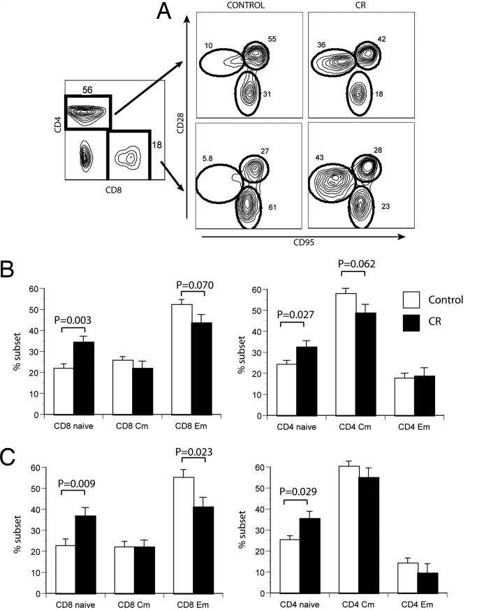
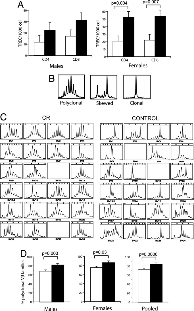
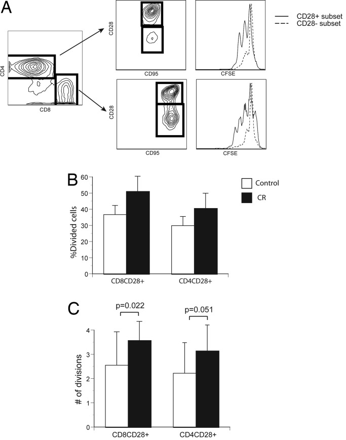
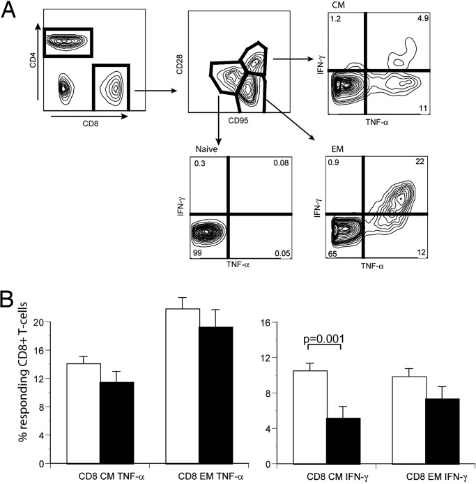
Caloric restriction (CR) has long been known to increase median and maximal lifespans and to decreases mortality and morbidity in short-lived animal models, likely by altering fundamental biological processes that regulate aging and longevity. In rodents, CR was reported to delay the aging of the immune system (immune senescence), which is believed to be largely responsible for a dramatic increase in age-related susceptibility to infectious diseases. However, it is unclear whether CR can exert similar effects in long-lived organisms. Previous studies involving 2- to 4-year CR treatment of long-lived primates failed to find a CR effect or reported effects on the immune system opposite to those seen in CR-treated rodents. Here we show that long-term CR delays the adverse effects of aging on nonhuman primate T cells. CR effected a marked improvement in the maintenance and/or production of naïve T cells and the consequent preservation of T cell receptor repertoire diversity. Furthermore, CR also improved T cell function and reduced production of inflammatory cytokines by memory T cells. Our results provide evidence that CR can delay immune senescence in nonhuman primates, potentially contributing to an extended lifespan by reducing susceptibility to infectious disease.


Conflict of interest statement

The authors declare no conflict of interest.

Figures

Fig. 1.
Fig. 1.
Phenotypic changes in aged RM T cells as a consequence of CR. (A) Example of four-color flow cytofluorometry analysis of PBMC from a representative pair of age-matched CON and CR animals. PBMC were stained with antibodies against CD8β, CD4, CD28, and CD95; were gated on CD8β or CD4 populations (Left); and were analyzed for the expression of CD28 and CD95 (Right). Circles denote N, CM, and EM subsets (encompassing >94% of all cells), as described in the text; numbers denote the percentage of cells in each population. (B and C) Cumulative percentages of T cell subsets were analyzed in all CON (open histograms) and CR (filled histograms) RM males (B) and females (C) over the four different analysis time points over a span of 42 months, as described in the text. Mean values are shown for each group at four time points, calculated from mean values at each time point ± SEM. Statistical analysis was performed using the mixed-effects model approach, and significance is indicated by two-sided P values above the histograms.
Fig. 2.
Fig. 2.
TCR repertoire analysis and TREC analysis of CON and CR T cells. (A) Estimation of recent thymic emigrants, using the TREC assay, was done as described in ref. . Results are shown as number of TREC per 103 T cells. (B) Representative examples of polyclonal (Left), skewed (Center), and clonal (Right) patterns of TCR CDR3 length polymorphism. (C) Example of CDR3 length analysis for all 24 TCRVβ families for representative age-matched CON and CR animals, performed as described in Materials and Methods. (D) Cumulative analysis (all animals) of CDR3 length polymorphisms in CON (open histograms) and CR (filled histograms) animals. For both A and D, statistical analysis was performed using the mixed-effects model.
Fig. 3.
Fig. 3.
Proliferation of CON- and CR-treated T cells in response to an agonistic antibody. (A) Illustration of flow cytofluorometry profiles of stimulated fluorescein (CFSE)-labeled CD4 and CD8 cells (stained and gated as described in Fig. 1) from a CON animal. Left shows CD4/CD8 profiles; Center shows CD28/95 profiles of gated cells from left; and Right depicts the successive fluorescein dilution in each proliferating subset at 96 h after stimulation. (B) Percentage of divided CD28+ T cells in CON (open histograms) and CR (filled histograms) males. Results are shown as mean ± SEM from a representative experiment out of three with comparable results. (C) Average number of divisions in the CD8+CD28+ and CD4+ CD28+ T cell subsets of CON (open histograms) or CR (filled histograms) male monkeys. All male animals were analyzed, and statistical analysis was as described in Fig. 1.
Fig. 4.
Fig. 4.
Production of proinflammatory cytokines by T cell subsets of CON and CR animals. (A) Illustration of flow cytofluorometry assay to measure ex vivo cytokine production by T cells. Blood T cells were stimulated for 6 h with an agonistic anti-TCR antibody, as described in Materials and Methods, and stained to reveal expression of CD4, CD8, CD95, CD28, and intracellular TNF-α. Selective gating is depicted by arrows and shows production of TNF-α by the N, CM, and EM subsets. (B) Cumulative analysis of TNF-α (Left) and IFN-γ (Right) in CD8 CM and EM T cell subsets of CON (open histograms) and CR (filled histograms) male monkeys. Statistical analysis was performed on all male animals as described in Fig. 1; significance is indicated by two-sided P values.

References

    1. McCay CM, Crowell MF, Maynard LA. Nutrition. 1989;5:155–171. discussion 172. - PubMed
    1. Masoro EJ. Mech Ageing Dev. 2005;126:913–922. - PubMed
    1. Heilbronn LK, Ravussin E. Am J Clin Nutr. 2003;78:361–369. - PubMed
    1. Hursting SD, Lavigne JA, Berrigan D, Donehower LA, Davis BJ, Phang JM, Barrett JC, Perkins SN. J Nutr. 2004;134:2482S–2486S. - PubMed
    1. Guarente L, Picard F. Cell. 2005;120:473–482. - PubMed

Publication types