. 2007 Jan;8(1):63-9.
doi: 10.1038/sj.embor.7400856. Epub 2006 Dec 8.

Structural and biochemical characterization of the yeast exosome component Rrp40

Affiliations

Structural and biochemical characterization of the yeast exosome component Rrp40

Anna Oddone et al. EMBO Rep. 2007 Jan.

Abstract

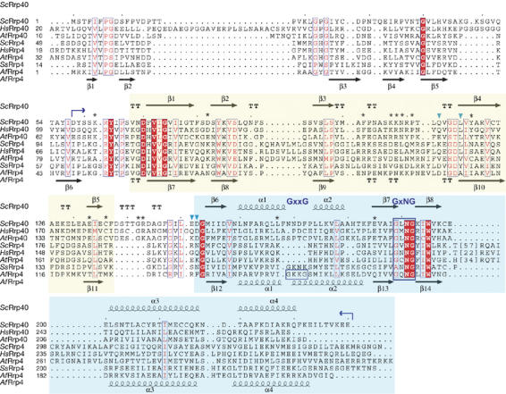
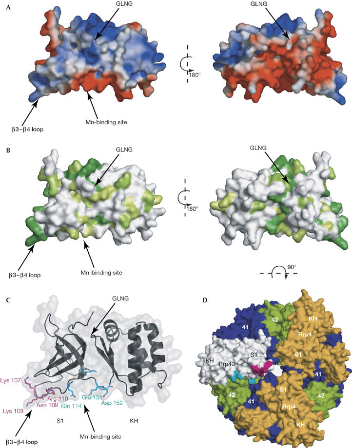
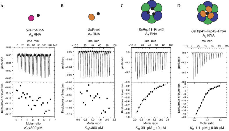
The exosome is a protein complex that is important in both degradation and 3'-processing of eukaryotic RNAs. We present the crystal structure of the Rrp40 exosome subunit from Saccharomyces cerevisiae at a resolution of 2.2 A. The structure comprises an S1 domain and an unusual KH (K homology) domain. Close packing of the S1 and KH domains is stabilized by a GxNG sequence, which is uniquely conserved in exosome KH domains. Nuclear magnetic resonance data reveal the presence of a manganese-binding site at the interface of the two domains. Isothermal titration calorimetry shows that Rrp40 and archaeal Rrp4 alone have very low intrinsic affinity for RNA. The affinity of an archaeal core exosome for RNA is significantly increased in the presence of the S1-KH subunit Rrp4, indicating that multiple subunits might contribute to cooperative binding of RNA substrates by the exosome.


Figures

Figure 1
Figure 1
Structure-based sequence alignment of Rrp40 and Rrp4 exosome subunits. Secondary structure elements in Saccharomyces cerevisiae Rrp40 (ScRrp40) and Archaeoglobus fulgidus Rrp4 (AfRrp4) are shown above and below the sequence alignment, respectively. The boundaries of Rrp40ΔN are indicated by blue arrows. The sequence alignment includes the sequences of Rrp40 and Rrp4 from S. cerevisiae, Homo sapiens (Hs), Arabidopsis thaliana (At), Sulfolobus solfataricus (Ss) and A. fulgidus. The ScRrp40 S1 and KH domain boundaries are indicated by yellow and light blue backgrounds, respectively. The canonical GxxG motif (present only in AfRrp4) and the exosome-specific GxNG sequence are boxed and indicated above the sequence. Stars indicate the residues conserved only in the Rrp40 family (but not in Rrp4). Blue arrowheads indicate residues affected in the Mn2+ titration. The alignment was obtained using SSM (http://www.ebi.ac.uk/msd-srv/ssm) and ProbCons (http://probcons.stanford.edu/).
Figure 2
Figure 2
Structure of Saccharomyces cerevisiae Rrp40ΔN. (A) Crystal structure of S. cerevisiae Rrp40ΔN (comprising residues 63–236). The S1 domain, the KH domain and the conserved GxNG motif are indicated. (B) Superposition of the structures of Rrp40ΔN (magenta) and of AfRrp4 (orange; Büttner et al, 2005). The two different GxxG sequence motifs present in AfRrp4 are indicated. (C) Detailed view of the GxNG loop connecting β7 and β8 in the ScRrp40 KH domain. Hydrogen bonds formed by Asn 191 with residues of the S1 domain are indicated by dotted lines. (D) View of the S1–KH domain interface. Conserved hydrophobic side chains, which stabilize the domain interface, are shown in black. Af, Archaeoglobus fulgidus.
Figure 3
Figure 3
Charge, conservation and interaction surfaces of Rrp40ΔN. Molecular surface representations of Rrp40: left, the same view as that depicted in Fig 2; right, rotated by 180° along a vertical axis. (A) Molecular surfaces are coloured blue and red according to positive and negative electrostatic potential, respectively. (B) The degree of sequence conservation among Rrp40 orthologues is mapped on the surface representation. Dark or light green indicates residues that are fully or partially conserved in Rrp40 orthologues, respectively (compare with Fig 1). (C) The conserved residues of the β3–β4 loop and the residues affected on addition of Mn2+ are shown in magenta and cyan, respectively. (D) Model of Rrp40ΔN in the context of the exosome, obtained by replacing one of the Rrp4 subunits in the structure of the Archaeoglobus fulgidus (Af) exosome by Rrp40ΔN. The conserved residues in the β3–β4 loop of the S1 domain and those affected by the addition of Mn2+ are shown in magenta and cyan, respectively. The AfRrp41 and AfRrp42 subunits are shown in blue and green, respectively, and the two AfRrp4 subunits are shown in orange. Ribbon and surface representations were generated with PyMOL (http://pymol.sourceforge.net).
Figure 4
Figure 4
RNA binding of exosome subunits studied by isothermal titration calorimetry. (A) Isothermal titration calorimetry (ITC) of yeast Rrp40ΔN (25 μM) with a solution of an A8 RNA oligonucleotide (670 μM). (B) ITC of SsRrp4 (20 μM) with a solution of A7 RNA (210 μM). (C) ITC of SsRrp41–Rrp42 (43 μM) with a solution of A7 RNA (430 μM). (D) ITC of SsRrp41–Rrp42–Rrp4 (14 μM) with a solution of A7 RNA (70 μM). For all titrations, the raw data are shown in the upper panel, and the integrated heat data, corrected for dilution, are shown in the lower panel. Above the ITC data is a schematic representation of the species in solution: magenta and orange circles represent ScRrp40 and SsRrp4 subunits respectively; blue and green circles represent Ss RNase PH-like subunits; small, black circles represent RNA molecules. Sc, Sulfolobus cerevisiae; Ss, Sulfolobus solfataricus.

References

    1. Allmang C, Kufel J, Chanfreau G, Mitchell P, Petfalski E, Tollervey D (1999) Functions of the exosome in rRNA, snoRNA and snRNA synthesis. EMBO J 18: 5399–5410 - PMC - PubMed
    1. Butler JS (2002) The yin and yang of the exosome. Trends Cell Biol 12: 90–96 - PubMed
    1. Büttner K, Wenig K, Hopfner KP (2005) Structural framework for the mechanism of archaeal exosomes in RNA processing. Mol Cell 20: 461–471 - PubMed
    1. Chekanova JA, Shaw RJ, Wills MA, Belostotsky DA (2000) Poly(A) tail-dependent exonuclease AtRrp41p from Arabidopsis thaliana rescues 5.8 S rRNA processing and mRNA decay defects of the yeast ski6 mutant and is found in an exosome-sized complex in plant and yeast cells. J Biol Chem 275: 33158–33166 - PubMed
    1. Delaglio F, Grzesiek S, Vuister G, Zhu G, Pfeifer J, Bax A (1995) NMRPipe: a multidimensional spectral processing system based on UNIX Pipes. J Biomol NMR 6: 277–293 - PubMed

Publication types

MeSH terms