. 2007 Jan;8(1):91-8.
doi: 10.1038/sj.embor.7400853. Epub 2006 Dec 8.

Pin1 stabilizes Emi1 during G2 phase by preventing its association with SCF(betatrcp)

Affiliations

Pin1 stabilizes Emi1 during G2 phase by preventing its association with SCF(betatrcp)

Cyril Bernis et al. EMBO Rep. 2007 Jan.

Abstract

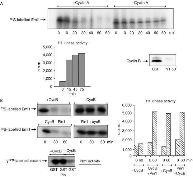
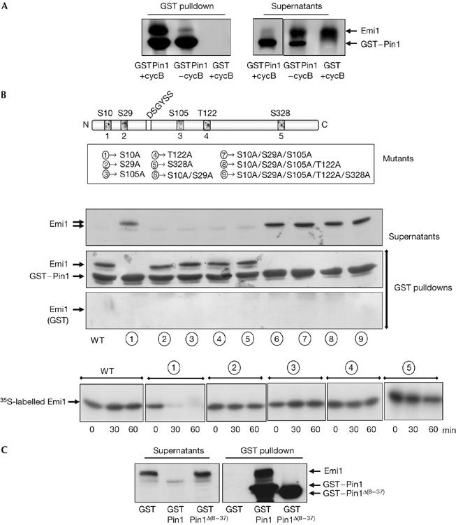
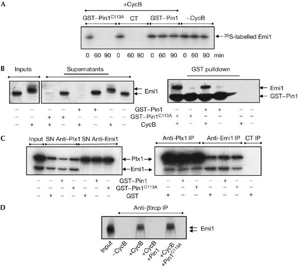
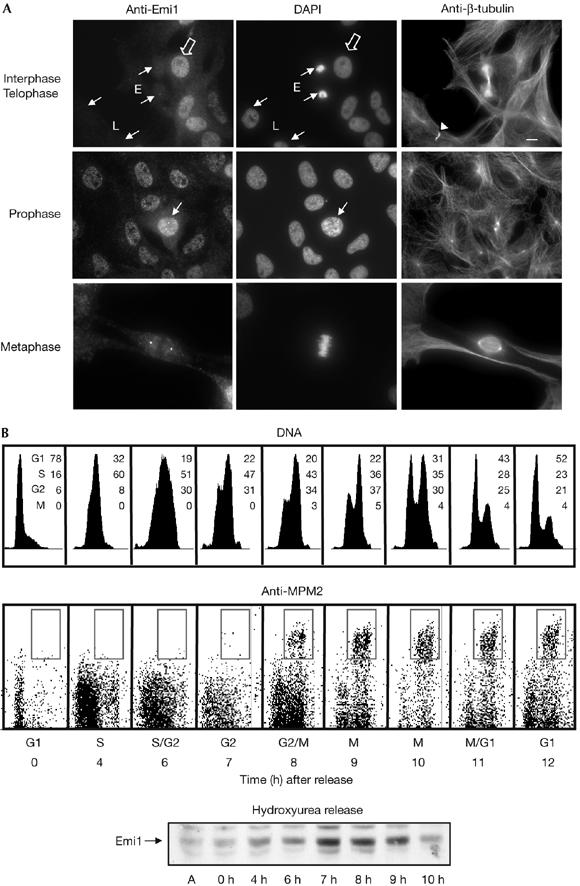
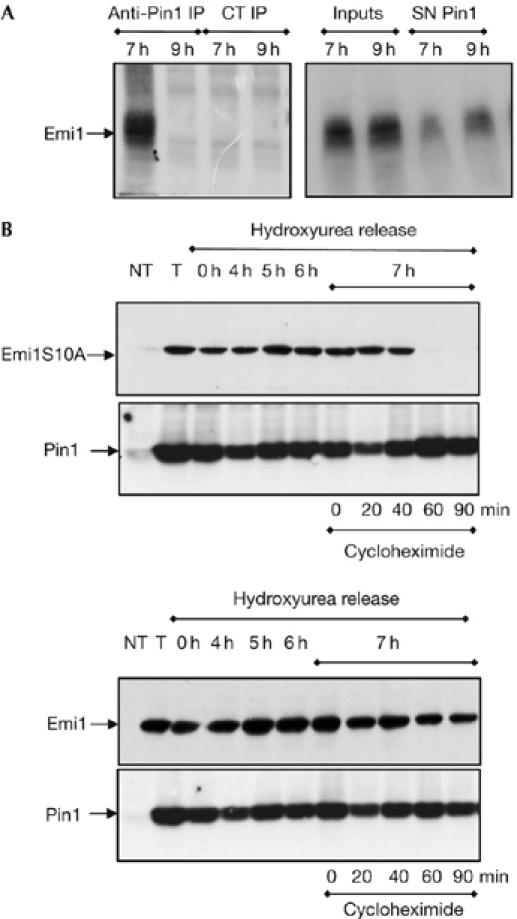
The anaphase-promoting complex (APC) early mitotic inhibitor 1 (Emi1) is required to induce S- and M-phase entries by stimulating the accumulation of cyclin A and cyclin B through APC(Cdh1/cdc20) inhibition. In this report, we show that Emi1 proteolysis can be induced by cyclin A/cdk (cdk for cyclin-dependent kinase). Paradoxically, Emi1 is stable during G2 phase, when cyclin A/cdk, Plx1 and SCF(betatrcp) (SCF for Skp1-Cul1-Fbox protein)--which play a role in its degradation--are active. Here, we identify Pin1 as a new regulator of Emi1 that induces Emi1 stabilization by preventing its association with SCF(betatrcp). We show that Pin1 binds to Emi1 and prevents its association with betatrcp in an isomerization-dependent pathway. We also show that Emi1-Pin1 binding is present in vivo in XL2 cells during G2 phase and that this association protects Emi1 from being degraded during this phase of the cell cycle. We propose that S- and M-phase entries are mediated by the accumulation of cyclin A and cyclin B through a Pin1-dependent stabilization of Emi1 during G2.


Figures

Figure 1
Figure 1
Pin1 stabilizes Emi1 in vitro. (A) Cyclin A/cdk promotes Emi1 degradation in interphase egg extracts. Human recombinant cyclin A (1.2 ng/μl) was added to interphase egg extracts devoid of endogenous cyclin A and cyclin B by the addition of cycloheximide (INT 50′). After 15 min, radiolabelled Emi1 was added and samples were taken at the indicated time points to analyse the 35S-labelled Emi1 levels and H1 kinase activity. A sample of 2 μl of a metaphase II-arrested extract (CSF) and the INT 50′ extract was immunoblotted with cyclin B2 antibody. (B) Pin1 stabilizes Emi1 in mitotic extracts. Interphase egg extracts were divided into four groups. One of these groups was not supplemented (−CycB). The second group was supplemented with recombinant GST-cyclin B (+CycB) only. The third group was first supplemented with GST–cyclin B and 15 min later with GST–Pin1 (CycB+Pin1). Finally, radiolabelled Emi1 was added in these three groups. In the fourth group, GST–Pin1 and radiolabelled Emi1 were simultaneously added and 15 min later GST–cyclin B was also added (Pin1+cycB). At the indicated time points after the addition of Emi1, samples of 2.8, 1 and 10 μl were taken to analyse the 35S-labelled Emi1 levels, H1 kinase activity and Polo kinase (Plx1) activity. Time 0 of the H1 kinase activity corresponds to the time at which Emi1 protein was added to the extract. Time 0 in the degradation assays corresponds to the time at which all the components of the mixture were present in the extract. cdk, cyclin dependent kinase; Emi1, early mitotic inhibitor 1; GST, glutathione S-transferase; Pin1, protein interacting with NIMA1.
Figure 2
Figure 2
Emi1 binding to Pin1 depends on the phosphorylation of Ser 10 of Emi1 and on the WW domain of Pin1. (A) Emi1-translated interphase egg extracts were supplemented with the proteasome inhibitor 4 hydroxy-5iodo-3-nitro-phenylacetyl-leucine-leucine vinylsulphone and with sepharose beads coupled to either GST–Pin1 (GSTPin1) or GST (GST) proteins. Thirty minutes later, GST–cyclin B was added (+cycB) or not (−cycB) to the mix and incubated for an additional 30 min. Finally, a GST pulldown was carried out. The precipitated samples and 1 μl of supernatants were analysed by western blot sequentially with an Emi1 antibody and a Pin1 antibody. (B) Five point mutants corresponding to the five putative cyclin B/cdk1 phosphorylation sites and double, triple, quadruple and quintuple mutants were constructed and their binding to GST–Pin1 or, as a control, to GST was analysed in the presence of GST–cyclin B by GST pulldown analysis, as described in (A). Degradation of simple mutants was analysed in interphase egg extract supplemented with wild-type GST–Pin1 and GST–cycB as described in Fig 1B. (C) GST pulldown analyses were developed as described in (A), in the presence of GST–cyclin B and of either wild-type GST–Pin1 (GST–Pin1) or GST–Pin1 deleted of the WW domain (GST–Pin1Δ(8–37)). cdk, cyclin dependent kinase; Emi1, early mitotic inhibitor 1; GST, glutathione S-transferase; Pin1, protein interacting with NIMA1; WT, wild type.
Figure 3
Figure 3
Pin1-dependent isomerization mediates stabilization of Emi1 by preventing its association with SCFβtrcp. (A) Interphase egg extracts were supplemented with either GST–Pin1 or the GST–Pin1C113A mutant and radiolabelled Emi1. After 15 min, GST–cyclin B was added. At the indicated time points, a sample was removed and analysed by autoradiography to measure levels of Emi1. (B) GST pulldowns were developed as described in Fig 2A except for the addition of either the wild-type (GST–Pin1) or the mutated (GST–Pin1C113A) proteins. Samples were analysed by western blot sequentially with an Emi1 antibody and subsequently a Pin1 antibody. (C) Radiolabelled Emi1 (15 μl) and Plx1-translated (5 μl) interphase egg extracts were mixed and supplemented first with 4 hydroxy-5iodo-3-nitro-phenylacetyl-leucine-leucine vinylsulphone (NLVS) and subsequently with GST, GST–Pin1 or the Pin1 mutant GST–Pin1C113A in the presence of cyclin B, as described in Fig 2A. The different mixtures were immunoprecipitated with Plx1 (Anti-Plx1 IP), Emi1 (Anti-Emi1 IP) or control antibodies (CT IP) and the immunoprecipitates were analysed by autoradiography. The input and the supernatants (SN) correspond to 1 μl of mixed extracts. (D) Emi1-translated extracts were supplemented with NLVS, GST, GST–Pin1 or GST–Pin1C113A, as described above, in the presence or absence of cyclin B. Subsequently, mixes were immunoprecipitated with βtrcp antibodies and the immunoprecipitates were subjected to western blot with the Emi1 antibody. The input corresponds to 1 μl of non-supplemented Emi1-translated extract. Emi1, early mitotic inhibitor 1; GST, glutathione S-transferase; Pin1, protein interacting with NIMA1; Plx1, polo-like Xenopus kinase 1.
Figure 4
Figure 4
Emi1 localization in Xenopus tadpole cells during cell cycle. (A) XTC cells were fixed with 3.7% formaldehyde, permeabilized with 0.1% Triton X-100 and blocked with PBS–5% fetal bovine serum. Subsequently, coverslips were incubated with Emi1 antibody (affinity purified, 5 μg/ml) and anti-β-tubulin (mAb, 1:10). Anti-mouse Alexa 488- and anti-rabbit Alexa 555-conjugated 2° antibodies (1/600 and 1/1,000, respectively; Molecular Probes, Leiden, The Netherlands) and 4,6-diamidino-2-phenylindole (10 μg/ml) were used. Small arrows indicate cells in early (E) and late (L) telophase as well as in prophase. The arrowhead points to midbody. The empty arrow shows cells in interphase. Scale bar, 10 μm. (B) Fluorescence-activated cell sorting analysis of synchronized XL2 cells. Cells synchronized by hydroxyurea block were collected at the indicated time points after release, labelled with MPM2 antibody and counterstained with propidium iodide. The percentage of cells in each cell cycle stage is indicated. Equal amounts of lysates from XL2 cells synchronized in (A) were taken at the indicated time points and used for immunoblotting to analyse endogenous Emi1 levels. Emi1, early mitotic inhibitor 1; XTC, Xenopus tadpole cells.
Figure 5
Figure 5
Pin1 associates and stabilizes Emi1 during G2 phase in XL2 cells. (A) Equal amounts of lysates from XL2 cells synchronized in Fig 4B were taken at the indicated time points and subjected to immunoprecipitation assay by using control (CT) or Pin1 antibodies. Immunoprecipitates (IP) as well as an equal amount of inputs and supernatants (SN) were immunoblotted with Emi1 antibody and are shown. (B) XL2 asynchronous cells were either not transfected (NT) or transfected (T, 0, 4, 5 and 6 h) with PCS2 wild type or S10A Emi1 as well as with PCS2-Pin1 vectors and synchronized by hydroxyurea block. Cells were lysed at 0, 4, 5 and 6 h after hydroxyurea release and the levels of the Emi1 and Pin1 ectopic proteins were analysed by western blot. At the 7 h release time point, cells were incubated with cycloheximide and lysed at the indicated time points after protein synthesis inhibition to evaluate wild-type and S10A Emi1 degradation patterns. The low endogenous Emi1 and Pin1 levels were not visualized at the low chemiluminescence exposure time used in this experiment to show the ectopic forms. Emi1, early mitotic inhibitor 1; Pin1, protein interacting with NIMA1.

References

    1. Castro A, Bernis C, Vigneron S, Labbe JC, Lorca T (2005) The anaphase-promoting complex: a key factor in the regulation of cell cycle. Oncogene 24: 314–325 - PubMed
    1. Eldridge AG, Loktev AV, Hansen DV, Verschuren EW, Reimann JD, Jackson PK (2006) The Evi5 oncogene regulates cyclin accumulation by stabilizing the anaphase-promoting complex inhibitor Emi1. Cell 27: 367–380 - PubMed
    1. Furuno N, den Elzen N, Pines J (1999) Human cyclin A is required for mitosis until mid prophase. J Cell Biol 147: 295–306 - PMC - PubMed
    1. Guardavaccaro D, Kudo Y, Boulaire J, Barchi M, Busino L, Donzelli M, Margottin-Goguet F, Jackson PK, Yamasaki L, Pagano M (2003) Control of meiotic and mitotic progression by the F box protein β-Trcp1 in vivo. Dev Cell 4: 799–812 - PubMed
    1. Hansen DV, Loktev AV, Ban KH, Jackson PK (2004) Plk1 regulates activation of the anaphase promoting complex by phosphorylating and triggering SCFβTrCP-dependent destruction of the APC inhibitor Emi1. Mol Biol Cell 15: 5623–5634 - PMC - PubMed

Publication types

MeSH terms