. 2007 Jan;9(1):25-35.
doi: 10.1038/ncb1514. Epub 2006 Dec 10.

H3K9 methylation and RNA interference regulate nucleolar organization and repeated DNA stability

Affiliations

H3K9 methylation and RNA interference regulate nucleolar organization and repeated DNA stability

Jamy C Peng et al. Nat Cell Biol. 2007 Jan.

Abstract

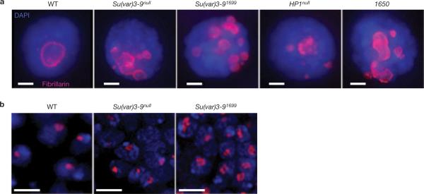
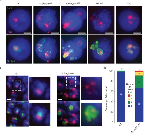
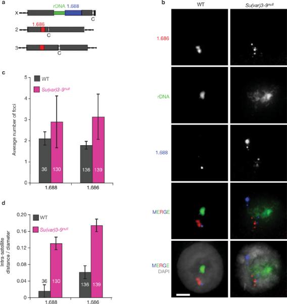
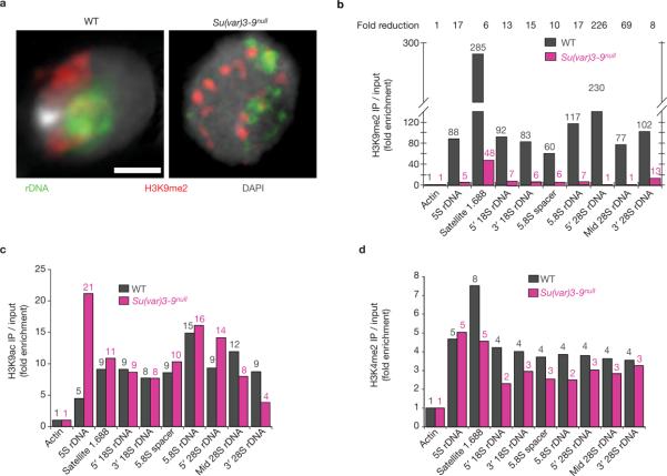
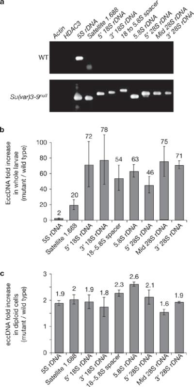
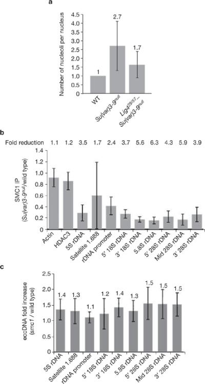
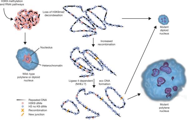
Investigations aimed at identifying regulators of nuclear architecture in Drosophila demonstrated that cells lacking H3K9 methylation and RNA interference (RNAi) pathway components displayed disorganized nucleoli, ribosomal DNA (rDNA) and satellite DNAs. The levels of H3K9 dimethylation (H3K9me2) in chromatin associated with repeated DNAs decreased dramatically in Su(var)3-9 and dcr-2 (dicer-2) mutant tissues compared with wild type. We also observed a substantial increase in extrachromosomal circular (ecc) repeated DNAs in mutant tissues. The disorganized nucleolus phenotype depends on the presence of Ligase 4 and ecc DNA formation is not induced by removal of cohesin. We conclude that the structural integrity and organization of repeated DNAs and nucleoli are regulated by the H3K9 methylation and RNAi pathways, and other regulators of heterochromatin-mediated silencing. In addition, repeated DNA stability involves suppression of non-homologous end joining (NHEJ) or other recombination pathways. These results suggest a mechanism for how local chromatin structure can regulate genome stability, and the organization of chromosomal elements and nuclear organelles.


Figures

Figure 1
Figure 1
Su(var) mutants contain multiple nucleoli. (a) Immunofluorescence microscopy with antibodies against the nucleolus marker fibrillarin (red) in whole-mount salivary-gland nuclei from wild-type (WT), Su(var)39null, Su(var)391699, HP1null and Su(TDA-PEV)1650 homozygous mutants. Wild-type cells have one nucleolus, whereas the mutants display multiple nucleoli. DAPI, blue. The scale bar represent 10 μm. (b) Immunofluorescence microscopy of fibrillarin in whole-mount imaginal disc and brain tissues from wild-type and Su(var)39 mutants are shown. The single, wild-type nucleolus tended to be round, whereas nucleoli in the mutants were irregular (lobed) and larger. Quantitative analysis showed that 44% of Su(var)39null mutant nuclei contained lobed nucleoli (n = 55), versus 10% for wild type (n = 51; P <0.001). The scale bars represent 5 μm.
Figure 2
Figure 2
Su(var) mutants have dispersed rDNA foci, each of which forms an ectopic nucleolus. (a) FISH of rDNA (red) and immunfluorescence microscopy of fibrillarin (green) were performed on whole-mount salivary glands from wild-type, Su(var)39null, Su(var)391699, HP1null and Su(TDA-PEV)1650 homozygous mutants. DAPI, blue. There is a single site of rDNA in >98% of wild-type nuclei, whereas the Su(var) mutant nuclei contain multiple rDNA foci, which are all surrounded by fibrillarin. The scale bars represent 15 μm. (b) Combined rDNA FISH (red) and immunofluoresence microscopy of fibrillarin (green) of whole-mount imaginal disc and brain tissues from wild-type and Su(var)39null mutant larvae. Wild-type nucleoli contain a single, compact rDNA focus, whereas Su(var)39null mutants frequently display multiple rDNA foci. The scale bars represent 3 μm. Boxed nuclei are shown at higher magnification to the right of each image. (c) Quantitative analysis of the number of rDNA foci in wild-type and Su(var)39null diploid nuclei. 98% of wild-type cells (n = 96) contain one rDNA signal, compared with 67% of Su(var)39null nuclei, and the percentages with two, three and four rDNA signals were 24%, 7% and 2%, respectively (average = 1.44 ± 0.73 rDNA foci per mutant nucleus, n = 53, P <0.001).
Figure 3
Figure 3
Satellite DNA organization is disrupted in Su(var)39null mutant nuclei. (a) Schematic representation of the locations of rDNA and satellite DNAs in the Drosophila melanogaster genome (not to scale). The rDNA is located in the heterochromatin of the X and Y sex chromosomes, the 1.688 satellite (359-bp repeats) is next to the X rDNA, and the 1.686 satellite is in the heterochromatin of chromosomes 2 and 3. (b) FISH was performed on whole-mount polytene salivary glands isolated from wild-type and Su(var)39null mutants. In wild-type nuclei, specific satellite DNAs are localized at single sites, and the different satellite signals are close to each other. In Su(var)39null mutant nuclei, individual satellite DNAs are dispersed to multiple sites and are not clustered with other satellites. DAPI, grey. FISH probe colours correspond to the diagram in a. The scale bars represent 15 μm. (c) The number of 1.688 and 1.686 foci were significantly higher in mutant nuclei compared with wild type (P <0.001). (d) Distances between satellite signals were quantified in three-dimensional reconstructions. The intra-satellite distances in Su(var)39null mutant nuclei were significantly higher than in wild type (P <0.001). The n values are indicated within the histograms and error bars correspond to s.d.
Figure 4
Figure 4
Analysis of histone modifications in chromatin containing repeated DNA in wild-type and Su(var)39null cells. (a) Immunofluorescence microscopy using antibodies that specifically bind H3K9me2 (red) combined with FISH for rDNA (green) in squashed diploid nuclei. In wild-type nuclei, rDNA chromatin partially overlaps with the H3K9me2 signals (correlation coefficient = 0.61 ± 0.08), and the amount of overlap was significantly reduced in Su(var)39null mutants (correlation coefficient = 0.40 ± 0.03; P <0.01). The scale bar represents 3 μm. (b) ChIP analysis of H3K9me2 levels in wild-type and Su(var)39null mutant imaginal disc tissues. The graph shows H3K9me2 levels for the repeated DNAs examined by PCR, standardized to actin and HDAC single-copy controls (see Methods); values are averages of five ChIP experiments and are shown above each bar. In wild-type cells, the 1.688 satellite (359-bp repeats), 5S rDNA (in chromosome 2 euchromatin) and the rDNA on the sex chromosomes contain significant enrichment for H3K9me2, compared with input chromatin and controls. H3K9me2 levels in chromatin derived from Su(var)39null mutant tissues were significantly reduced (6–226-fold) compared with wild type. (c, d) ChIP analysis of two modifications associated with active or open chromatin (H3K9ac and H3K4me2). Small enrichment for these modifications was observed on repeated DNAs in wild-type chromatin, compared with input and single-copy controls. For most of the repeated DNAs, levels were not significantly altered in Su(var)39null mutant chromatin (P >0.5 for all regions). H3K9ac levels were significantly increased in 5S rDNA in the mutants (P <0.05), and H3K4me2 was significantly decreased for the 1.688 satellite (P <0.05). Values are averages of two experiments and are shown above each bar.
Figure 5
Figure 5
Levels of extrachromosomal repeated DNAs are significantly increased in Su(var)39null mutant tissues compared with wild type. (a) Extrachromosomal DNA was isolated from wild-type and Su(var)39null mutant larvae using the Hirt supernatant method, and PCR reactions, terminated at logarithmic phase of amplification, were performed to evaluate the amounts of eccDNA corresponding to specific sequences (see Methods). The gel shows an example of the PCR reactions for the specific regions examined. EccDNAs from the single-copy genes (actin and HDAC3) were not detected in either wild-type or mutant larvae. The asterisk indicates that the band in the 1.688 satellite lane corresponds to the primers, not the PCR products. (b) Quantification demonstrates that the amount of eccDNA for the 1.688 satellite and different regions of the rDNA are significantly higher in Su(var)39null mutants compared with wild-type (20–78-fold enrichment); the increase for 5S rDNA was only twofold because wild-type larvae contain high levels of ecc 5S rDNA. The values, as shown above each bar, are averages of three sample extractions. (c) Quantification of PCR products indicates that the amount of eccDNA in Su(var)39null mutant diploid cells is about twofold higher than in wild type. The values, shown above each bar, were averages of three sample extractions, and P values were <0.05 for the regions examined. The error bars correspond to s.d.
Figure 6
Figure 6
The RNAi pathway is also required to maintain the structural integrity of repeated DNAs and the nucleolus. (a) Combined rDNA FISH (red) and immunofluorescence microscopy of fibrillarin (green) shows that dcr-2L811fsx polytene nuclei contain multiple rDNA foci and ectopic nucleoli. (b) The histogram shows the average number of nucleoli in different RNAi mutants examined (absolute values are indicated above each bar). Mutations at all loci contained significantly more nucleoli than wild type (P <0.001, except aubQC42, P <0.004). The hlsdel215 allele of SpnE1 had a mild phenotype (P = 0.083). (c) ChIP analysis reveals reduced H3K9me2 levels in dcr-2L811fsx chromatin compared with wild type (P <0.05), more so for rDNA than the 5S rDNA and satellite 1.688. Values are averages of four PCR reactions from two ChIP experiments and the overall fold reductions are shown above. (d) Immunofluorescence microscopy for H3K9me2 (red) and fibrillarin (green) on whole-mount brains and imaginal discs from wild-type and dcr-2L811fsx mutants. H3K9me2 localizes predominantly in DAPI-bright heterochromatin regions in wild-type nuclei, but is more broadly distributed in dcr-2L811fsx nuclei. The scale bar represents 5 μm. (e) ecc rDNA levels in dcr-2L811fsx mutant larvae are significantly higher than in wild type (13–29-fold increases), but eccDNA levels for 5S rDNA and satellite 1.688 did not increase. The values indicated above each bar are averages of two PCR experiments from two sample extractions (P <0.05). The error bars correspond to s.d.
Figure 7
Figure 7
Effects of Ligase 4 and cohesin mutants on ectopic nucleolus and eccDNA formation. (a) Ligase 4 mutations partially suppress ectopic nucleolus formation in Su(var)39 mutants. Average nucleolus number of Lig4nullSu(var)3–9null polytene nuclei is 1.7 (n = 83), which is significantly lower (P <0.001) than the average 2.7 (n = 54) nucleoli observed in Su(var)39null mutant nuclei. Absolute values are indicated above each bar. (b) ChIP analysis shows reduced SMC1 levels at repeated DNAs in Su(var)39null chromatin, relative to wild type; fold reductions are shown (P <0.05 for all repeated DNA except 5S rDNA). Values are averages of four PCR reactions from two ChIP experiments. (c) The amount of eccDNA from satellite 1.688 and rDNA in smc1exc46l mutant tissues do not differ significantly from wild type. Absolute values are indicated above each bar. Error bars indicate s.d.
Figure 8
Figure 8
A schematic representation of a model for regulation of nuclear architecture by the H3K9 methylation and RNAi pathways. In wild-type diploid and polytene nuclei, the majority of the heterochromatin contains H3K9me2 and a single nucleolus forms around the rDNA. Loss of H3K9me2 from repeated DNA, due to Su(var)39, HP1 or RNAi mutations, causes chromatin decondensation and elevated recombination between repeated DNA copies. The recombination process results in formation of eccDNAs that localize throughout the nucleoplasm, causing dispersal of satellite DNAs (not shown) and, in the case of rDNA, the formation of ectopic nucleoli. Decondensation is proposed to be primarily responsible for the lobed structure of rDNA and nucleoli in diploid cells, with a minor contribution from low levels of ecc rDNA formation (dotted line). In polytene cells, decondensation is likely to be a prerequisite for increased recombination, but the much higher levels of ecc rDNA is proposed to generate the majority of the ectopic nucleoli.

Comment in

  • Heterochromatin: condense or excise.
    Pikaard C, Pontes O. Pikaard C, et al. Nat Cell Biol. 2007 Jan;9(1):19-20. doi: 10.1038/ncb0107-19. Nat Cell Biol. 2007. PMID: 17199128 No abstract available.

Similar articles

Cited by

References

    1. Francastel C, Schubeler D, Martin DI, Groudine M. Nuclear compartmentalization and gene activity. Nature Rev. Mol. Cell Biol. 2000;1:137–143. - PubMed
    1. Cremer T, Cremer C. Chromosome territories, nuclear architecture and gene regulation in mammalian cells. Nature Rev. Genet. 2001;2:292–301. - PubMed
    1. John B. The biology of heterochromatin. In: Verma RS, editor. Heterochromatin: Molecular and Structural Aspects. Cambridge University Press; Cambridge: 1988. pp. 1–147.
    1. Martin C, Zhang Y. The diverse functions of histone lysine methylation. Nature Rev. Mol. Cell Biol. 2005;6:838–849. - PubMed
    1. Jenuwein T, Allis CD. Translating the histone code. Science. 2001;293:1074–1080. - PubMed

Publication types