. 2007 Jan;117(1):206-17.
doi: 10.1172/JCI29293. Epub 2006 Dec 7.

The type III TGF-beta receptor suppresses breast cancer progression

Affiliations

The type III TGF-beta receptor suppresses breast cancer progression

Mei Dong et al. J Clin Invest. 2007 Jan.

Abstract

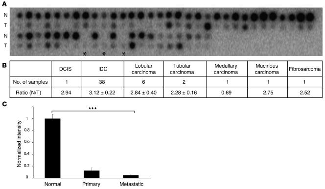
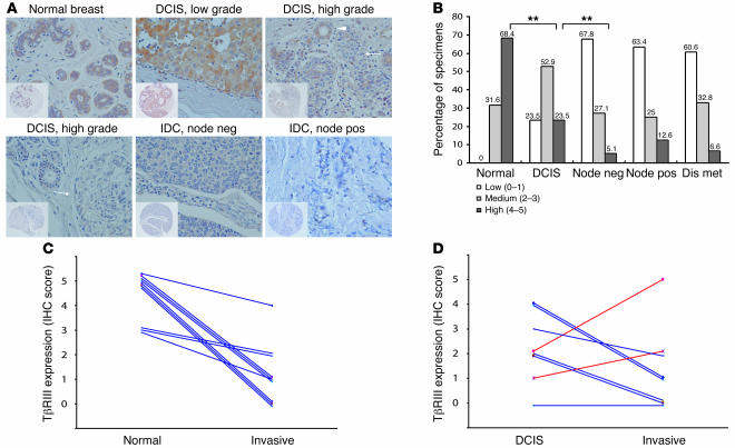
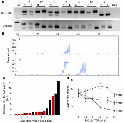
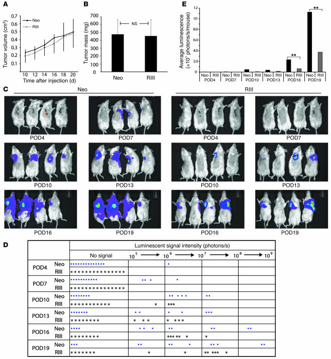
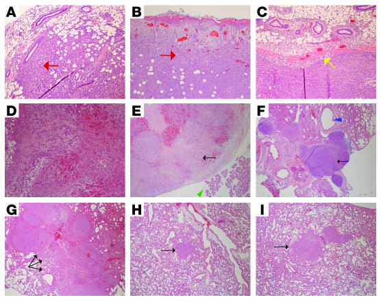
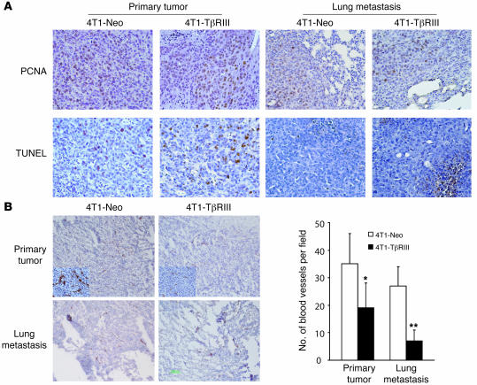
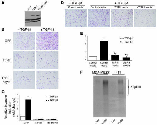
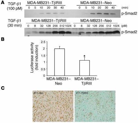
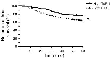
The TGF-beta signaling pathway has a complex role in regulating mammary carcinogenesis. Here we demonstrate that the type III TGF-beta receptor (TbetaRIII, or betaglycan), a ubiquitously expressed TGF-beta coreceptor, regulated breast cancer progression and metastasis. Most human breast cancers lost TbetaRIII expression, with loss of heterozygosity of the TGFBR3 gene locus correlating with decreased TbetaRIII expression. TbetaRIII expression decreased during breast cancer progression, and low TbetaRIII levels predicted decreased recurrence-free survival in breast cancer patients. Restoring TbetaRIII expression in breast cancer cells dramatically inhibited tumor invasiveness in vitro and tumor invasion, angiogenesis, and metastasis in vivo. TbetaRIII appeared to inhibit tumor invasion by undergoing ectodomain shedding and producing soluble TbetaRIII, which binds and sequesters TGF-beta to decrease TGF-beta signaling and reduce breast cancer cell invasion and tumor-induced angiogenesis. Our results indicate that loss of TbetaRIII through allelic imbalance is a frequent genetic event during human breast cancer development that increases metastatic potential.


Figures

Figure 1
Figure 1. Loss of TβRIII mRNA expression during mammary carcinogenesis.
(A) TβRIII mRNA levels were detected by hybridizing [32P]-labeled human TβRIII cDNA probe to the Clontech Cancer Profiling Array I. The portion of the array containing breast samples is shown, with tumor specimens (T) and matched normal breast tissue (N). Asterisks indicate metastatic specimens corresponding to the normal and tumor samples spotted on the immediate left. (B) Quantitative data were obtained by analyzing the array with NIH ImageJ software, summarized as the ratio relative to normal breast, and expressed as mean ± SEM. (C) Quantitative data from matched normal, primary breast tumor, and metastatic breast tumor tissue expressed as mean ± SEM. ***P < 0.0001, ANOVA.
Figure 2
Figure 2. Progressive loss of TβRIII protein expression during mammary carcinogenesis.
(A) Representative IHC analysis of TβRIII expression (original magnification, ×40) in normal breast ductal cells, in different grades of DCIS, and in lymph node–negative (node neg) and –positive (node pos) IDC. Insets depict staining of entire tissue core (original magnification, ×10). Immunoreactivity for TβRIII was scored as 0–5 and categorized as low (0–1), medium (2–3), or high (4–5). Note the absence of TβRIII staining in IDC and high-grade DCIS (arrows) versus presence of staining in normal ducts and normal-appearing ducts adjacent to the DCIS lesion (arrowhead). (B) Summary of IHC results, with percentages shown. Dis met, distant metastasis. **P < 0.01, 2-tailed Fisher’s exact probability. (C) Patient-matched normal and invasive breast cancer IHC TβRIII scores. (D) Patient-matched DCIS and invasive breast cancer IHC TβRIII scores.
Figure 3
Figure 3. Frequent LOH of the TGFBR3 gene locus in human breast cancers correlates with loss of TβRIII mRNA expression.
LOH analysis was performed on DNA extracted from 26 human breast cancer specimens and matching normal lymphocytes. (A) Representative results showing allelic loss in tumors 1, 2, and 6 (denoted by asterisks) when PCR products were separated on a MetaPhor agarose gel. Microsatellite markers D1S1588 and D1S188 are described in Methods. (B) LOH was confirmed using an ABI sequencer and quantified using GeneScan software. A representative sample with LOH is shown. (C) Quantitative real-time PCR analysis of TβRIII mRNA levels in breast cancer specimens with (red bars) and without (black bars) LOH. (D) Quantitative real-time PCR analysis of mRNA levels of TβRI, TβRII, and TβRIII in MDA-MB231 cells in response to TGF-β1 (100 pM) stimulation for the indicated times.
Figure 4
Figure 4. TβRIII delayed and decreased metastatic potential of breast cancer cells in vivo.
Either 4T1-Neo (Neo) or 4T1-TβRIII (RIII) cells (75,000 cells/mouse) were implanted into the axillary mammary fat pads of BALB/c mice. (A) Primary tumor growth was recorded by measuring tumor size every 2 days beginning at 10 days after injection and presented as mean ± SEM. (B) Weight of the primary tumors upon surgical removal on day 20 after injection. Data are mean ± SEM (n = 16). (C) Bioluminescence imaging was performed every 3 postoperative days (POD). Representative images are shown. Red and violet signals correspond to the maximum and minimum intensity values, respectively, with other colors representing the values in between. (D) Record of luminescent signals for every mouse in each group at the indicated time points. (E) Average luminescent signal in each group at the indicated time points. **P < 0.01.
Figure 5
Figure 5. TβRIII decreased tumor cell invasiveness and metastasis in vivo.
Representative H&E staining (original magnification, ×10) of (A and B) primary tumors from mice implanted with 4T1-Neo cells exhibiting local invasion (red arrows) of tumor cells into the adjacent normal mammary tissue (A) and skin (B); (C) a representative primary tumor from mice implanted with 4T1-TβRIII cells demonstrating the absence of local invasion, as indicated by the clear margin between the tumor and the adjacent normal mammary tissue (yellow arrow); (D) a recurring tumor in a mouse at the primary injection site of 4T1-Neo cells exhibiting internal bleeding due to invasion of tumor cells into the blood vessels; (E) a metastatic tumor (black arrow) adjacent to the pancreas (green arrowhead) found on the mesentery of a mouse implanted with 4T1-Neo cells; (F) a significantly enlarged paratracheal lymph node adjacent to the trachea (blue arrowhead) containing metastatic tumor cells (black arrow) in a mouse with 4T1-Neo cells, indicating the presence of lymphatic metastasis; (G) multiple large metastatic tumor nodules (black arrows) in the lung of a mouse implanted with 4T1-Neo cells; and (H and I) representative lung metastases in mice implanted with 4T1-TβRIII cells (black arrows).
Figure 6
Figure 6. TβRIII inhibits tumor angiogenesis without altering cancer cell proliferation and apoptosis in vivo.
(A) Tissue sections of primary tumors and lung metastases from mice implanted with 4T1-Neo and 4T1-TβRIII cells were immunostained for PCNA and TUNEL to evaluate cell proliferation and apoptosis, respectively. Representative staining frequency and intensity is shown (original magnification, ×40). (B) Immunostaining of CD31 (original magnification, ×10) was performed as a marker to evaluate angiogenesis. Note the decreased number and size of tumor-associated blood vessels as well as decreased staining intensity (insets; original magnification, ×100) in 4T1-TβRIII primary tumors and lung metastases. Values are the averages from 6 mice and expressed as mean ± SD. *P < 0.05; **P < 0.01.
Figure 7
Figure 7. Restoration of TβRIII expression inhibits Matrigel invasiveness of MDA-MB231 breast cancer cells.
(A) MDA-MB231 cells were infected with equivalent amounts of adenoviral constructs carrying GFP, HA-tagged TβRIII, and a TβRIII mutant lacking the entire cytoplasmic domain (TβRIIIΔcyto). Expression of the transgenes was confirmed by Western blotting of cell lysate using anti-HA antibody. (B and C) Matrigel invasion assay. Adenovirally infected MDA-MB231 cells (75,000 cells) were seeded in a Matrigel-coated upper chamber and treated with TGF-β1 (15 pM) 2 hours later. Cell invasion through the Matrigel after 24 hours’ incubation was detected by H&E staining and quantitated. (D and E) Matrigel invasion assay was performed after resuspending MDA-MB231 cells in the conditioned media collected from pcDNA3.1-Neo–, TβRIII-, and sTβRIII-transfected COS-7 cells. Data are mean ± SEM, n = 3 in triplicate. **P < 0.01. (F) Detection of sTβRIII in media of MDA-MB231–TβRIII and 4T1-TβRIII cells by [125I]TGF-β1–binding crosslinking followed by immunoprecipitation.
Figure 8
Figure 8. TβRIII attenuates Smad2 phosphorylation in vitro and in vivo.
(A) TβRIII-overexpressing and control MDA-MB231 cells were treated with TGF-β1 under the indicated conditions, and cell lysates were analyzed with a phospho-Smad2 (p-Smad2) antibody. (B) Cells were transfected with pE2.1 and pSVβ vector. Luciferase activity was determined after 24 hours of TGF-β1 treatment (100 pM) and is expressed as the fold induction over no TGF-β treatment after adjusting for β-galactosidase expression. This assay was performed in triplicate at least 3 times. *P < 0.05. (C) Phosphorylated Smad2 immunostaining of tissue sections from 4T1-Neo and 4T1-TβRIII primary tumors. Representative results are shown. Note the significant decrease in staining intensity in the 4T1-TβRIII tumor. Original magnification, ×40.
Figure 9
Figure 9. Low levels of TβRIII predict decreased recurrence-free survival in women with breast cancer.
Five-year recurrence-free survival for breast cancer with high or low TβRIII expression was analyzed based on a microarray data set containing 286 patients. *P < 0.05.

References

    1. Massague J. TGF-beta signal transduction. Annu. Rev. Biochem. 1998;67:753–791. - PubMed
    1. Jhappan C., et al. Targeting expression of a transforming growth factor beta 1 transgene to the pregnant mammary gland inhibits alveolar development and lactation. Embo J. 1993;12:1835–1845. - PMC - PubMed
    1. Pierce D.F., et al. Inhibition of mammary duct development but not alveolar outgrowth during pregnancy in transgenic mice expressing active TGF-beta 1. Genes Dev. 1993;7:2308–2317. - PubMed
    1. Pierce D.F., et al. Mammary tumor suppression by transforming growth factor beta 1 transgene expression. Proc. Natl. Acad. Sci. U. S. A. 1995;92:4254–4258. - PMC - PubMed
    1. Decensi A., et al. Correlation between plasma transforming growth factor-beta 1 and second primary breast cancer in a chemoprevention trial. Eur. J. Cancer. 1998;34:999–1003. - PubMed

Publication types

MeSH terms

LinkOut - more resources