Case Reports
. 2007 Jan;80(1):186-94.
doi: 10.1086/510499. Epub 2006 Nov 15.

The Meckel-Gruber syndrome gene, MKS3, is mutated in Joubert syndrome

Affiliations
Case Reports

The Meckel-Gruber syndrome gene, MKS3, is mutated in Joubert syndrome

Lekbir Baala et al. Am J Hum Genet. 2007 Jan.

Abstract

Joubert syndrome (JS) is an autosomal recessive disorder characterized by cerebellar vermis hypoplasia associated with hypotonia, developmental delay, abnormal respiratory patterns, and abnormal eye movements. The association of retinal dystrophy and renal anomalies defines JS type B. JS is a genetically heterogeneous condition with mutations in two genes, AHI1 and CEP290, identified to date. In addition, NPHP1 deletions identical to those that cause juvenile nephronophthisis have been identified in a subset of patients with a mild form of cerebellar and brainstem anomaly. Occipital encephalocele and/or polydactyly have occasionally been reported in some patients with JS, and these phenotypic features can also be observed in Meckel-Gruber syndrome (MKS). MKS is a rare, autosomal recessive lethal condition characterized by central nervous system malformations (typically, occipital meningoencephalocele), postaxial polydactyly, multicystic kidney dysplasia, and ductal proliferation in the portal area of the liver. Since there is obvious phenotypic overlap between JS and MKS, we hypothesized that mutations in the recently identified MKS genes, MKS1 on chromosome 17q and MKS3 on 8q, may be a cause of JS. After mutation analysis of MKS1 and MKS3 in a series of patients with JS (n=22), we identified MKS3 mutations in four patients with JS, thus defining MKS3 as the sixth JS locus (JBTS6). No MKS1 mutations were identified in this series, suggesting that the allelism is restricted to MKS3.


Figures

Figure  1.
Figure  1.
Pathological features of fetal patients with MKS3 mutations. A, Histological pattern of kidney and liver (hematoxylin-eosin-safran staining) in fetuses JS-661 (at 28 WG) and JS-660 (at 30 WG). Kidney is shown at low (panels a and d) and high (panels b and e) magnification. Liver is shown at high magnification (panels c and f). Kidney histology shows conserved corticomedullary organization with 6 to 8 generations of mature glomerules. Microcysts are found in the deep cortex in JS-661. Tubular microcysts in medulla (black arrows) are observed in both. Liver histology shows a portal fibrosis with important and diffuse bile-duct proliferation (blue arrows) in both fetuses. B, Neuropathological findings of brainstem and cerebellum. Transversal section of the brainstem and cerebellum at the level of cerebellar peduncles in fetus JS-660 (panel g) compared with a control individual (panel h). In panel g, note the inverted molar tooth aspect due to a dysmorphic fourth ventricle (V4) flanked laterally by thick cerebellar peduncles (P). The roof of the fourth ventricle is formed by the remnant of vermian structure (V). Cerebellar folia contain heterotopic Purkinje cells (data not shown). The hypoplastic pons longitudinal tracts (LT) have a chaotic organization, and the transverse fibers are reduced (panel i), as compared with a control individual (panel j); this is better seen on a caudal level with higher magnification.
Figure  2.
Figure  2.
Brain MRI of a normal control individual, an affected girl aged 7 years (JS-09), and an affected girl aged 14 years (JS-05) carrying MKS3 mutations. A, Sagittal MRI in JS-09 shows a dilated fourth ventricle slightly superiorly displaced (white arrows). The fourth ventricle in JS-05 was normally sized but superiorly displaced. Both patients JS-09 and JS-05 show a superior vermian dysplasia. The middle and inferior segments of the vermis are hypoplastic. B, Axial images at the level of the cerebellar hemispheres show that the middle and inferior segments of the vermis are hypoplastic in patients JS-09 and JS-05; the two hemispheres are not separated by any vermian structure. Patient JS-09 had also an hemispheric cerebellar dysplasia. C, Axial images showing the MTS (white arrow) at the level of the superior cerebellar peduncles in patients JS-09 and JS-05.
Figure  3.
Figure  3.
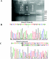
Analysis of two MKS3 splice-site mutations at the RNA level. A, RT-PCR with primers located in MKS3 exons 20 (forward) and 26 (reverse) in the father of patient JS-05, JS-05, and a control individual. Expected wild-type size (754 bp) is seen in the control individual, whereas in patient JS-05 the smaller band (617 bp) corresponds to the transcript without exon 23. The father of patient JS-05 shows both normal and mutated transcripts. B, Direct sequencing of the RT-PCR of patient JS-05 shows a skipping from exons 22 to 24. C, Direct sequencing of the RT-PCR of patient JS-09 showing the wild-type (upper line) and abnormal (lower line) transcripts corresponding to the insertion of 16 intronic bp (boxed).

References

Web Resources

    1. Online Mendelian Inheritance in Man (OMIM),http://www.ncbi.nlm.nih.gov/Omim/ (for JS, MKS, and BBS) - PubMed
    1. PolyPhen, http://genetics.bwh.harvard.edu/pph/
    1. Splice Site Prediction by SSF, http://www.umd.be/SSF
    1. TMHMM Server, http://www.cbs.dtu.dk/services/TMHMM/

References

    1. Joubert M, Eisenring JJ, Andermann F (1968) Familial dysgenesis of the vermis: a syndrome of hyperventilation, abnormal eye movements and retardation. Neurology 18:302–303 - PubMed
    1. Patel S, Barkovich AJ (2002) Analysis and classification of cerebellar malformations. AJNR Am J Neuroradiol 23:1074–1087 - PMC - PubMed
    1. Ferland RJ, Eyaid W, Collura RV, Tully LD, Hill RS, Al-Nouri D, Al-Rumayyan A, Topcu M, Gascon G, Bodell A, et al (2004) Abnormal cerebellar development and axonal decussation due to mutations in AHI1 in Joubert syndrome. Nat Genet 36:1008–101310.1038/ng1419 - DOI - PubMed
    1. Sayer JA, Otto EA, O’Toole JF, Nurnberg G, Kennedy MA, Becker C, Hennies HC, Helou J, Attanasio M, Fausett BV, et al (2006) The centrosomal protein nephrocystin-6 is mutated in Joubert syndrome and activates transcription factor ATF4. Nat Genet 38:674–68110.1038/ng1786 - DOI - PubMed
    1. Valente EM, Brancati F, Silhavy JL, Castori M, Marsh SE, Barrano G, Bertini E, Boltshauser E, Zaki MS, Abdel-Aleem A, et al (2006) AHI1 gene mutations cause specific forms of Joubert syndrome-related disorders. Ann Neurol 59:527–53410.1002/ana.20749 - DOI - PubMed

Publication types

LinkOut - more resources