. 2007 Jan 26;1130(1):54-66.
doi: 10.1016/j.brainres.2006.10.072. Epub 2006 Dec 11.

Developmental and injury-induced expression of alpha1beta1 and alpha6beta1 integrins in the rat spinal cord

Affiliations

Developmental and injury-induced expression of alpha1beta1 and alpha6beta1 integrins in the rat spinal cord

K Adam Baker et al. Brain Res. .

Abstract

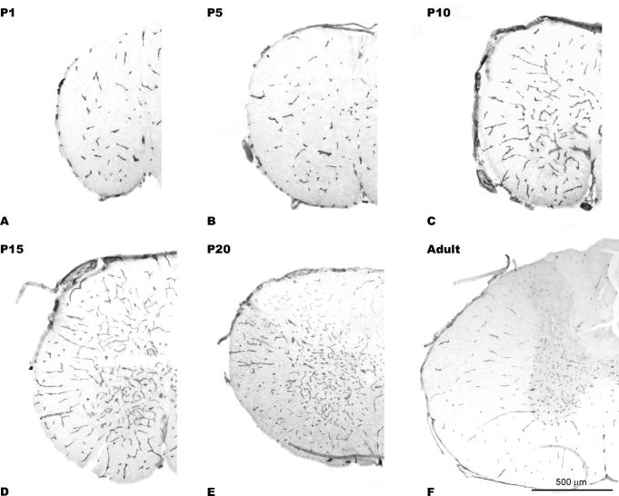
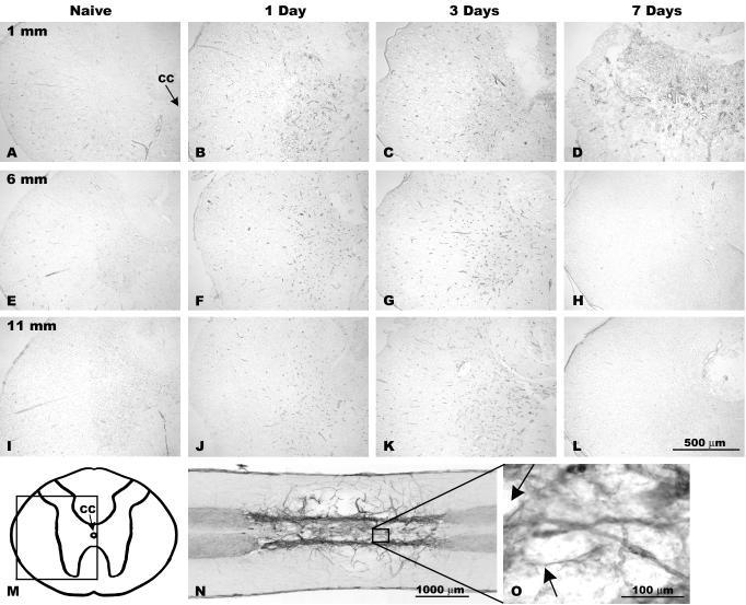
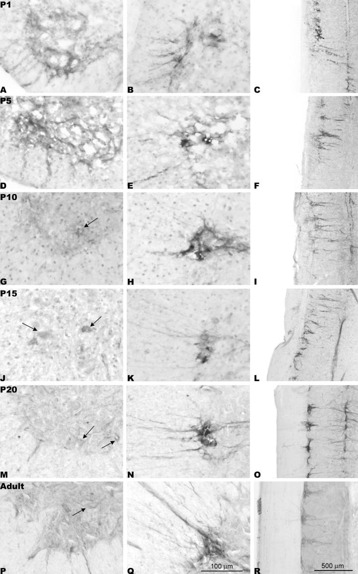
Loss and damage to blood vessels are thought to contribute to secondary tissue loss after spinal cord injury. Integrins might be therapeutic targets to protect the vasculature and/or promote angiogenesis, as their activation can promote tubule formation and survival of endothelial cells in vitro. Here, we show that immunostaining with an antibody against the alpha1beta1 integrin heterodimer is present only in blood vessels from postnatal day 1 (P1) through adulthood in Sprague-Dawley rats. After a spinal cord contusion at T9 in adults, the area of alpha1beta1 integrin positive blood vessels increases within 11 mm from the injury site at 3 days post-injury and remains prominent within the injured core only at 7 days. Staining for the alpha6beta1 integrin heterodimer increases in blood vessels between P10 and adulthood and is present in preganglionic neurons of the intermediolateral cell column (IML) at all ages. The alpha6beta1 integrin is also expressed by motor neurons postnatally, and oligodendrocyte precursors (OPCs), as previously reported. After the contusion, the area of alpha6beta1-stained blood vessels is increased at 3 days and most prominently, 1 mm from the injury site, followed by a significant reduction at 7 days, when alpha6beta1 integrin staining is most prominent around the injured core. Staining is also present in a subset of microglia and/or macrophages. These results raise the possibility that alpha1beta1 and alpha6beta1 integrins in blood vessels might be targeted to reduce blood vessel loss and promote angiogenesis, which may promote tissue sparing after spinal cord injury.


Figures

Figure 1
Figure 1
Expression of α1β1 integrin in the developing spinal cord. α1β1-positive blood vessels are readily observed at all developmental ages in transverse sections through the thoracic spinal cord. The number of blood vessels and complexity of the vasculature gradually increases through P1 (A), P5 (B), P10 (C), P15 (D) and P20 (E) spinal cord with vessels appearing to be thickest at P15. α1β1 immunostaining can also be observed on the surface of the spinal cord, likely representing menigeal fibroblasts.
Figure 2
Figure 2
Expression of α1β1 by blood vessels in the naïve and injured spinal cord. (A-C) α1β1 expression is specific to SMI-71-positive blood vessels in naïve animals. (D-E) Thin fibrous α1β1-positive, SMI-71-negative ‘streamers’ (arrows) can be seen within the lesion cavity and may represent small immature vessels not yet possessing a mature blood-spinal cord barrier, and thus not yet expressing SMI-71.
Figure 3
Figure 3
α1β1 integrin positive blood vessels increase after thoracic spinal cord contusions. (A, E, I) Few blood vessels are detected by α1β1 immunostaining in sham-operated animals. The α1β1 integrin staining appears to increase at 1 (B, F, J) and 3 days (C, G, K) after contusion up to 11 mm from 1 mm of the injury epicenter. At 7 days, increased integrin expression is seen 1 mm from the injury epicenter prominently within the injured core (D) but appears similar to baseline levels at 6 (H) and 11 mm (L). M) Schematic indicating the location of the panels in Fig. 3A-L and Fig. 8A-L. N) A horizontal section demonstrating α1β1 integrin staining in the spinal cord 7 days after injury. Many α1β1-positive blood vessels are seen extending primarily rostrocaudally through the lesion site with vessels extending radially from these vessels into the lateral white matter. (O) A higher magnification of vessels within the lesion cavity demonstrates the thin α1β1 integrin-positive processes often seen attached to blood vessels at this time-point following injury (arrows). CC = central canal.
Figure 4
Figure 4
Integrin-positive blood vessel increases after spinal cord injury: quantification. (A) The percentage of α1β1-immunopositive blood vessel area increases up to 3 days after injury within 11 mm of the injury epicenter. However, at 7 days an increase in area is only seen at 1 mm, with decreases below that of sham-operated animals being seen at 6 and 11 mm. (B) An increase in the percentage of α6β1-positive blood vessel area is seen 1-3 days after injury up to 11 mm from the epicenter, whereas at 7 days the percent blood vessel area is similar to that of sham-operated animals at 1 mm and at other distances the blood vessel area drops below that of sham-operated animals.
Figure 5
Figure 5
Expression of α6β1 integrin in the developing spinal cord. From P10 (D) to adulthood (F) staining of blood vessels with α6β1 was seen (arrows). In P1 (A), P5 (B) and P10 (C) transverse sections of the spinal cord, α6β1 staining was also seen within preganglionic cell somata within the intermediolateral cell column (IML) and associated processes (arrowheads) motor neurons in the ventral horn and their processes.
Figure 6
Figure 6
α6β1 expression within the ventral horn and IML of the rat spinal cord. Left and middle columns: higher magnification images of Figure 4. Left column: expression of α6β1 integrin is highest within ventral horn motor neurons at P1 (A) and P5 (D). Also note the α6β1-positive processes extending from the motor neurons towards the ventrolateral spinal cord (the transition zone to the ventral root) at this age. Expression of α6β1 integrin is reduced but still evident in motor neurons at later ages (arrows). The middle column shows transverse sections and right column horizontal sections (rostral is at the top, midline at the right): α6β1-positive preganglionic neurons of the intermediolateral nucleus are seen at each developmental age. Furthermore, α6β1-positive dendrites can be seen as early as P1 (B,C) extending medially with fewer processes projecting laterally. At P10 (E,F) α6β1-positive processes appear to project also rostrocaudally within the IML cell column and at the midline. The complexity of the α6β1-positive dendrites greatly increases at P20 (R,S) with intense staining seen within dendrites ventral to the central canal. A fragment of α6β1-positive meninges can be seen in (R).
Figure 7
Figure 7
Blood vessels, oligodendrocyte precursors and a subset of activated microglia have α6β1 integrin immunostaining in the adult rat spinal cord. A-C) In the normal spinal cord, α6β1 integrin is present in SMI-71-positive blood vessels (arrows) in normal spinal cord. Note that the α6β1-positive processes from the intermediolateral (IML) cell column (arrowheads) do not co-express SMI-71, suggesting that they are neuronal processes and not blood vessels. D-F) NG2-positive oligodendrocyte precursors also have α6β1 integrin immunostaining. G-I) GFAP-positive astrocytes do not have α6β1 integrin staining. These IML processes (arrowhead) are intimately intertwined with the astrocytic processes within the white matter. J-L) Resting microglia in the normal spinal cord also did not exhibit α6β1 integrin staining. M-O) A subset of Iba1-positive microglia/macrophages (arrows) express α6β1 integrin, 7 days after spinal cord contusion around the epicenter.
Figure 8
Figure 8
α6β1 integrin-positive blood vessels increase after thoracic spinal cord contusions. (A, E, I) Sham-operated rats demonstrated low levels of α6β1 in blood vessels and clear staining of IML preganglionic neurons and their processes. After spinal contusions, α6β1 staining appeared to be increased in blood vessels at 1 and 3 days, 1 mm rostral to the injury site (B and C, respectively) and at 6 (F and G, respectively) and 11 mm (J and K, respectively). (D, H, L) At 7 days, α6β1 integrin staining patterns approached normal levels. The same labeling pattern was seen in sections caudal to the lesion.

References

    1. Andreasen SO, Thomsen AR, Koteliansky VE, Novobrantseva TI, Sprague AG, de Fougerolles AR, Christensen JP. Expression and functional importance of collagen-binding integrins, α1β1 and α2β1, on virus-activated T cells. J. Immunol. 2003;171:2804–2811. - PubMed
    1. Babic AM, Kireeva ML, Kolesnikova TV, Lau LF. CYR61, a product of a growth factor-inducible immediate early gene, promotes angiogenesis and tumor growth. Proc. Natl. Acad. Sci. U.S.A. 1998;95:6355–6360. - PMC - PubMed
    1. Baker KA, Hagg T. An adult rat spinal cord contusion model of sensory axon degeneration: a preconditioning lesion or the estrus cycle do not affect outcome. J. Neurotrauma. 2005;22:415–428. - PubMed
    1. Bartholdi D, Rubin BP, Schwab ME. VEGF mRNA induction correlates with changes in the vascular architecture upon spinal cord damage in the rat. Eur. J. Neurosci. 1997;9:2549–2560. - PubMed
    1. Ben-Horin S, Bank I. The role of very late antigen-1 in immune-mediated inflammation. Clin. Immunol. 2004;113:119–129. - PubMed

Publication types

MeSH terms