. 2007 Feb 9;366(1):244-57.
doi: 10.1016/j.jmb.2006.11.036. Epub 2006 Nov 11.

Structural analysis of the plakin domain of bullous pemphigoid antigen1 (BPAG1) suggests that plakins are members of the spectrin superfamily

Affiliations

Structural analysis of the plakin domain of bullous pemphigoid antigen1 (BPAG1) suggests that plakins are members of the spectrin superfamily

Julius J Jefferson et al. J Mol Biol. .

Abstract

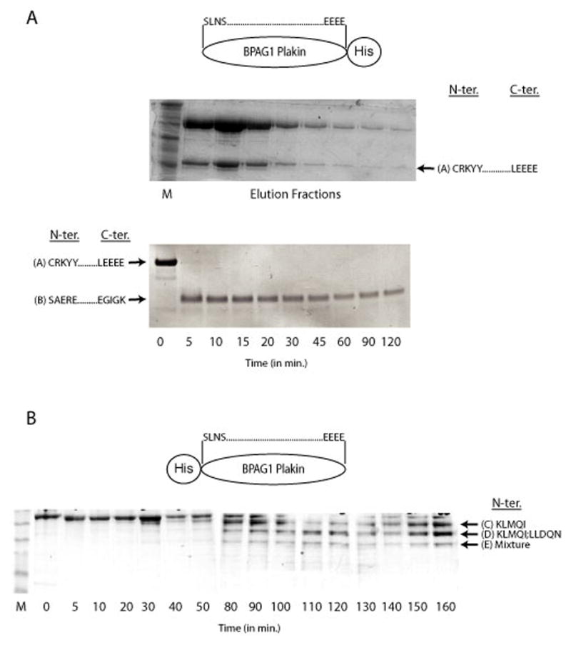
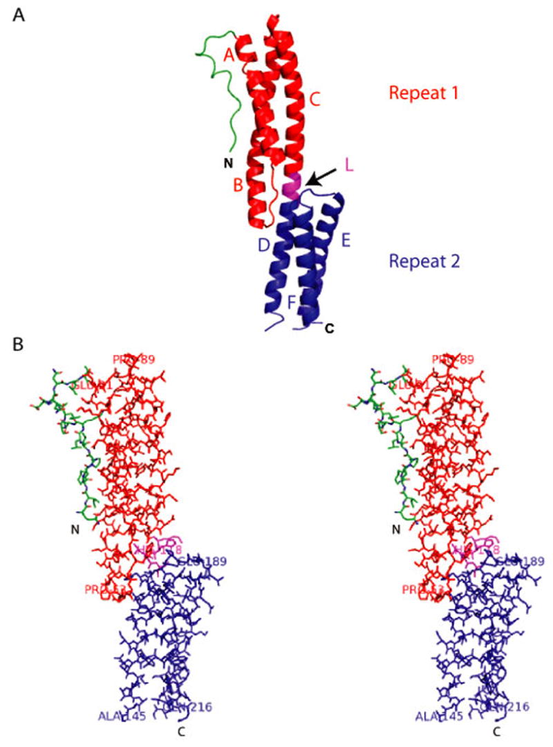
Bullous pemphigoid antigen 1 (BPAG1) is a member of the plakin family of proteins. The plakins are multi-domain proteins that have been shown to interact with microtubules, actin filaments and intermediate filaments, as well as proteins found in cellular junctions. These interactions are mediated through different domains on the plakins. The interactions between plakins and components of specialized cell junctions such as desmosomes and hemidesmosomes are mediated through the so-called plakin domain, which is a common feature of the plakins. We report the crystal structure of a stable fragment from BPAG1, residues 226-448, defined by limited proteolysis of the whole plakin domain. The structure, determined by single-wavelength anomalous diffraction phasing from a selenomethionine-substituted crystal at 3.0 A resolution, reveals a tandem pair of triple helical bundles closely related to spectrin repeats. Based on this structure and analysis of sequence conservation, we propose that the architecture of plakin domains is defined by two pairs of spectrin repeats interrupted by a putative Src-Homology 3 (SH3) domain.


Figures

Figure 1
Figure 1. (A) Purification of fragments of the C-terminal His-tagged Plakin Domain of BPAG1
SDS-PAGE of the final step of purification of the C-terminal tagged plakin domain of BPAG1 also yields a proteolytic fragment referred to as A (top gel). Limited proteolysis of fragment A by trypsin produced another fragment, referred to as B (bottom gel). The boundaries of the fragments based on N-terminal sequencing results and mass spectrometry are also shown. (B) Limited Proteolysis of the N-terminal His-tagged Plakin Domain of BPAG1. SDS-PAGE of a time course of a limited Proteinase K digest of the plakin domain of BPAG1with an N-terminal His-tag is shown. The first five N-terminal residues of the various fragments (C and D) are also shown. D contained 2 distinct peptides, whereas E consisted of a mixture of peptides whose identities could not be assigned with certainty. (C) Primary Sequence of the Plakin Domain of BPAG1. The N- and C-terminal amino acids identified in (A) and (B) are indicated in bold. The fragment that produced crystals is underlined. All the fragments used for crystallization experiments are enclosed in brackets and also summarized in Table 1. The first pair of identified spectrin repeats (Repeat 1 and 2), the predicted repeats (Repeat 3 and 4) and the putative SH3 domain are boxed. The alpha-helical regions designated as Z,Y,X,W and V based on the nomenclature of the plakin domain of desmoplakin by Green and colleagues; are also illustrated (arrows).
Figure 2
Figure 2. Structure of the Spectrin Repeats Identified within the Plakin Domain of BPAG1. (A)
A cartoon representation of the structure showing a loop-like region (in green) at the extreme N-terminus followed by a pair of spectrin repeats (Repeat 1 in red and Repeat 2 in blue) in tandem connected by a linker region (L in magenta) that is also helical in nature. The structure consists of helices A, B and C (Repeat 1) and helices D, E and F (Repeat 2). (B) Stereo diagram showing all atoms of the structure shown in (A).
Figure 3
Figure 3. Comparison of Spectrin Repeats of BPAG1 and Spectrin. (A)
Amino-acid sequence alignment of spectrin repeats (Repeat 1 and Repeat 2) identified within the plakin domain of BPAG1 with the representative spectrin repeats of known structure. The residues involved in the stacking interactions are shown (arrows). Conserved residues are highlighted in red and boxed in blue. SR-Spectrin repeat; HuEry-Human Erythroid; ChBr-Chicken Brain; AlphaAct-Alpha Actinin. (B) Stereoview of a superposition of spectrin repeat 1 of BPAG1 (red) with repeat 16 of chicken brain α-spectrin (blue). Rmsd=1.49Å for 91 alpha carbon atoms. (C) Inter-helical stacking interactions that are highly conserved in spectrin repeats involving a residue in each of the three helices constituting a repeat is illustrated.
Figure 4
Figure 4. BPAG1 and Other Plakins
Amino-acid sequence alignment of the fragment of BPAG1 (NT-BP-PKN) that produced crystals with other plakins is shown. The N-terminal region that forms a loop like structure is not conserved except with human BPAG1 where the overall conservation is high (>90%). The hydrophobic residues that form stacking interactions to stabilize the repeat are highly conserved in most plakins (arrows). Conserved residues are highlighted in red and boxed in blue. h-Homo sapiens; m-Mus musculus.
Figure 5
Figure 5. Comparison of Spectrin Repeats Within and Outside of the Plakin Domain of BPAG1
Amino-acid sequence alignment of the two spectrin repeats (R1 and R2) identified within the plakin domain of BPAG1 and the other spectrin repeats predicted outside the plakin domain of larger isoforms such as BPAG1a and BPAG1b. Conserved residues are highlighted in red and boxed in blue. Also included are additional repeats (R3 and R4) predicted within the plakin domain. Overall, there are 4 spectrin repeats in BPAG1e and 32 spectrin repeats in BPAG1a and BPAG1b. SR-spectrin repeat. The number following the prefix, SR, indicates the position of the spectrin repeat in sequential order.
Figure 6
Figure 6. Putative SH3 domain of BPAG1. (A)
Model of the SH3 domain of BPAG1 (in red; on top left) is shown. Superposition (on top right) of the crystal structure of SH3 domain of alpha-spectrin (in green) with the model of the SH3 domain of BPAG1 shows that the RT loop is larger in alpha-spectrin due to the presence of three extra residues. (B) Structure based amino-acid sequence alignment of the putative SH3 domain of BPAG1 with equivalent regions from other plakins, the SH3 domain of alpha-spectrin and representative Src family of proteins. The five anti-parallel beta-strands (β-1 to β-5) forming the SH3 domain of alpha-spectrin are indicated (horizontal arrows on top). The five residues important for interactions, based on the structural analysis of the complexes between SH3 domain and proline-rich ligands, are also shown (vertical arrows at the bottom). m-Mus musculus
Figure 7
Figure 7. (A) Nomenclature of the Plakin Domain
The spectrin repeats identified within the plakin domain of BPAG1 are shown in the context of the old nomenclature of the plakin domain of desmoplakin , which were predicted to consist of a series of alpha helical bundles termed - Z, Y, X, W and V. The pair of repeats includes the entire Z domain and most of the Y domain. Also shown is the proposed domain architecture of the entire plakin domain of BPAG1, based on the crystal structure reported in this study and domain prediction algorithms. (B) Domain Architecture of Plakins and Spectrins. A schematic of the major isoforms of BPAG1 and the alpha- and beta- subunits of erythroid spectrin is shown to highlight the similarities between the plakin and spectrin family of proteins. PRD-plakin repeat domains; ABD-actin binding domain; MTBD-microtubule binding domain. Whereas the amino-terminal ends are similar consisting of either the ABD or spectrin repeats in both families, the carboxyl-terminal ends are divergent. Plakins consist of domains that can interact directly with either microtubules or intermediate filaments extending their cytolinker functions to all the three major cytoskeletal networks in the cell. (C) Evolutionary Relationship Between Plakins and Spectrins. The phylogenetic tree of plakins and spectrins based on the percent identity of the first pair of spectrin repeats is shown. Members of the plakin family included are BPAG1 and its close homologue MACF, plakins found in desmosomes and hemidesmosomes, plectin and desmoplakin and those present in the cornified envelope of skin, periplakin and envoplakin. Shortstop and VAB10 are plakin family orthologs found in Drosophila melanogaster and Caenorhabditis elegans, respectively. Members of the spectrin family included are alpha-actinin, alpha- and beta- chains of erythroid spectrin, dystrophin, utrophin and nesprin. d-Drosophila melanogaster; h-Homo sapiens; m-Mus musculus

Similar articles

Cited by

References

    1. Jefferson JJ, Leung CL, Liem RK. Plakins: goliaths that link cell junctions and the cytoskeleton. Nat Rev Mol Cell Biol. 2004;5:542–53. - PubMed
    1. Leung CL, Green KJ, Liem RK. Plakins: a family of versatile cytolinker proteins. Trends Cell Biol. 2002;12:37–45. - PubMed
    1. Leung CL, Zheng M, Prater SM, Liem RK. The BPAG1 locus: Alternative splicing produces multiple isoforms with distinct cytoskeletal linker domains, including predominant isoforms in neurons and muscles. J Cell Biol. 2001;154:691–7. - PMC - PubMed
    1. Choi HJ, Park-Snyder S, Pascoe LT, Green KJ, Weis WI. Structures of two intermediate filament-binding fragments of desmoplakin reveal a unique repeat motif structure. Nat Struct Biol. 2002;9:612–20. - PubMed
    1. Getsios S, Huen AC, Green KJ. Working out the strength and flexibility of desmosomes. Nat Rev Mol Cell Biol. 2004;5:271–81. - PubMed

Publication types

MeSH terms

Associated data