

The neural correlates of feature-based selective attention when viewing spatially and temporally overlapping images
- PMID: 17161441
- PMCID: PMC1876667
- DOI: 10.1016/j.neuropsychologia.2006.10.019
The neural correlates of feature-based selective attention when viewing spatially and temporally overlapping images
Abstract
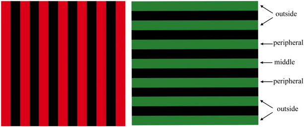
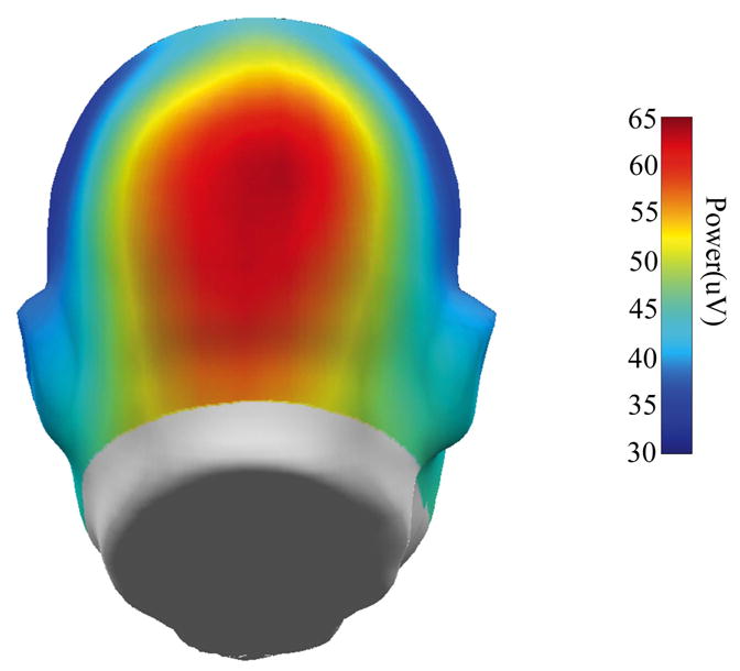
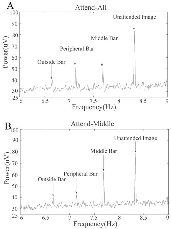
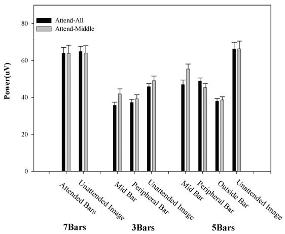
We used dense-array EEG to study the neural correlates of selective attention to specific features of objects that spatially overlapped an unattended image. Participants viewed superimposed images (horizontal and vertical bars differing in color) and attended to one image to identify bar width changes in specific locations. Images were frequency tagged so attention directed to unique parts of the stimuli could be tracked. Steady-state visual evoked potentials were used to quantify attention-related neural activity. As expected, selectively attending to specific parts of the attended image enhanced brain activity related to the attended element, and left unchanged activity elicited by spatially overlapping unattended stimuli. Under specific conditions, however, we found increased activity to unattended stimuli. The specificity of the selective attention effects presented herein, however, may be limited under certain complex stimulus conditions.
Figures
Similar articles
-
Normal electrocortical facilitation but abnormal target identification during visual sustained attention in schizophrenia.J Neurosci. 2008 Dec 10;28(50):13411-8. doi: 10.1523/JNEUROSCI.4095-08.2008. J Neurosci. 2008. PMID: 19074014 Free PMC article.
-
Objects are highlighted by spatial attention.J Cogn Neurosci. 2006 Feb;18(2):298-310. doi: 10.1162/089892906775783642. J Cogn Neurosci. 2006. PMID: 16494688
-
Attentional Selection of Feature Conjunctions Is Accomplished by Parallel and Independent Selection of Single Features.J Neurosci. 2015 Jul 8;35(27):9912-9. doi: 10.1523/JNEUROSCI.5268-14.2015. J Neurosci. 2015. PMID: 26156992 Free PMC article.
-
Strategic control of attention to objects and locations.J Neurosci. 2008 Jan 16;28(3):564-5. doi: 10.1523/JNEUROSCI.4386-07.2008. J Neurosci. 2008. PMID: 18199757 Free PMC article. Review. No abstract available.
-
Steady-State Visually Evoked Potentials and Feature-based Attention: Preregistered Null Results and a Focused Review of Methodological Considerations.J Cogn Neurosci. 2021 Apr;33(4):695-724. doi: 10.1162/jocn_a_01665. Epub 2021 Jan 8. J Cogn Neurosci. 2021. PMID: 33416444 Free PMC article. Review.
Cited by
-
Bottom-up attention orienting in young children with autism.J Autism Dev Disord. 2014 Mar;44(3):664-73. doi: 10.1007/s10803-013-1925-5. J Autism Dev Disord. 2014. PMID: 23996226 Free PMC article.
-
Visuocortical changes during delay and trace aversive conditioning: evidence from steady-state visual evoked potentials.Emotion. 2013 Jun;13(3):554-61. doi: 10.1037/a0031323. Epub 2013 Feb 11. Emotion. 2013. PMID: 23398582 Free PMC article.
-
The effects of attention pre-allocation and target-background integration on object-based attention.PLoS One. 2015 Mar 19;10(3):e0119414. doi: 10.1371/journal.pone.0119414. eCollection 2015. PLoS One. 2015. PMID: 25789772 Free PMC article.
-
Cross multivariate correlation coefficients as screening tool for analysis of concurrent EEG-fMRI recordings.J Neurosci Res. 2018 Jul;96(7):1159-1175. doi: 10.1002/jnr.24217. Epub 2018 Feb 6. J Neurosci Res. 2018. PMID: 29406599 Free PMC article.
-
Attention cueing in rivalry: insights from pupillometry.eNeuro. 2022 Jun 3;9(3):ENEURO.0497-21.2022. doi: 10.1523/ENEURO.0497-21.2022. Online ahead of print. eNeuro. 2022. PMID: 35667847 Free PMC article.
References
Publication types
MeSH terms
Grants and funding
LinkOut - more resources
Full Text Sources
Research Materials
