. 2007 Feb 23;144(4):1436-46.
doi: 10.1016/j.neuroscience.2006.11.002. Epub 2006 Dec 11.

Rapid neuroadaptation in the nucleus accumbens and bed nucleus of the stria terminalis mediates suppression of operant responding during withdrawal from acute opioid dependence

Affiliations

Rapid neuroadaptation in the nucleus accumbens and bed nucleus of the stria terminalis mediates suppression of operant responding during withdrawal from acute opioid dependence

S H Criner et al. Neuroscience. .

Abstract

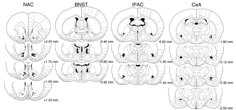
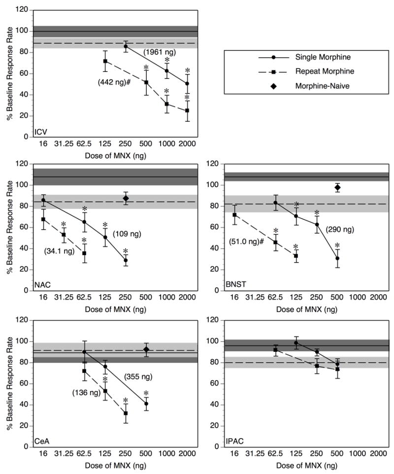
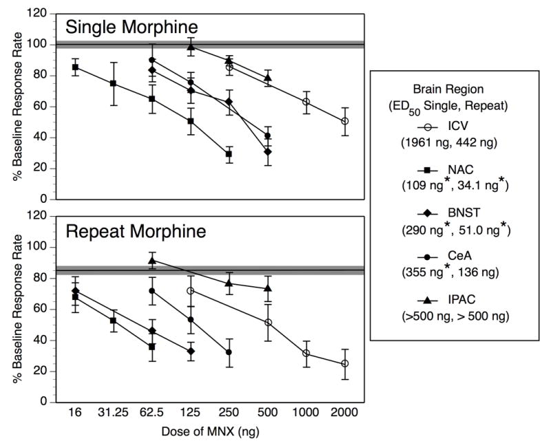
Single injections of morphine induce a state of acute opioid dependence in humans and animals, measured as precipitated withdrawal when an antagonist is administered 4-24 h after morphine. Additional morphine exposure at daily or weekly intervals results in further increases in withdrawal severity, suggesting that acute opioid dependence reflects the early stages in the development of a chronic state of dependence. The current study evaluated the role of the nucleus accumbens (NAC), bed nucleus of stria terminalis (BNST), interstitial nucleus of posterior limb of the anterior commissure (IPAC), and central amygdala (CeA) in the expression of antagonist-precipitated suppression of operant responding for food as a measure of withdrawal from acute opioid dependence. Rats trained on a fixed-ratio 15 schedule received one or four daily injections of morphine, with the lipophobic opioid antagonist methylnaloxonium (16-2000 ng) infused into one of the brain regions or the lateral ventricle (i.c.v.) 4 h after the final morphine injection. After acute morphine methylnaloxonium was more potent upon infusion into the NAC (17.9-fold potency shift), BNST (6.8-fold) and CeA (5.5-fold) than it was upon i.c.v. administration. Following repeat morphine the NAC and BNST but not CeA continued to show greater sensitivity relative to i.c.v. infusion (12.9-, 8.7-, and 3.2-fold potency shifts, respectively). The IPAC was insensitive to methylnaloxonium after acute or repeat morphine at doses that reliably suppressed responding upon i.c.v. infusion (125-500 ng). Thus, among the components of extended amygdala examined in this study, rapid neuroadaptation within the nucleus accumbens and bed nucleus of the stria terminalis appear to play the most prominent role in antagonist-precipitated suppression of operant responding during the early stages in the development of opioid dependence.


Figures

Fig. 1
Fig. 1
Cannula tip placements for the two highest doses tested under Single Morphine (left side) and Repeat Morphine (right side) conditions within each brain region examined. Values represent AP distance from bregma (mm) according to the atlas of Paxinos and Watson (1998). To maintain clarity of individual tip placements, cannula locations for other doses are not shown, but the range of tip placements illustrated is representative of all dose groups.
Fig. 2
Fig. 2
Following an acute treatment with 5.6 mg/mg morphine (Single Morphine, circles + solid lines) MNX dose-dependently suppresses operant responding for food after injection into the lateral ventricle (ICV), nucleus accumbens (NAC), bed nucleus of the stria terminalis (BNST) or central amygdala (CeA), but not the interstitial nucleus of the posterior limb of the anterior commissure (IPAC) (*p < 0.05 vs. vehicle-infused group under the corresponding morphine treatment condition (Single compared to Single, Repeat to Repeat). After 4 morphine treatments at daily intervals (Repeat Morphine, squares + dotted lines), the magnitude of response suppression elicited by MNX was further potentiated with ICV, and intra-BNST infusions, but not with intra-NAC or intra-CeA infusion (#p < 0.05, significant shift in MNX dose-effect function from Single to Repeat Morphine condition according to relative potency analysis, see Table 2 for further details of this analysis). In Morphine-Naive subjects (diamonds), MNX at the highest dose tested within active brain regions (NAC, BNST, CeA) failed to suppress operant responding relative to vehicle-infused controls. Individual data points on the MNX dose-effect functions represent mean ± SEM percent of baseline response rate. Values in parentheses are the ED50 values derived for the corresponding dose-response function. The solid horizontal line represents the mean of the group infused with vehicle 4 hr after Single Morphine Treatment, and the dotted horizontal line represents the mean of the Repeat Morphine vehicle-infused group; the shaded region around each horizontal line represents ± 1 SEM. N = 8–11/dose group.
Fig. 3
Fig. 3
This Figure re-presents the data from Fig. 2 in a way that facilitates comparison of MNX dose-effect functions across injection sites. Following a Single Morphine treatment (upper panel), MNX potency is significantly greater in the nucleus accumbens (NAC), bed nucleus of the stria terminalis (BNST), and central amygdala (CeA) than it is following intracerebroventricular infusion (ICV), but MNX potency in the interstitial nucleus of the posterior limb of the anterior commissure (IPAC) does not differ from ICV infusion. With Repeat Morphine treatment (lower panel), MNX remains significantly more potent in the NAC and BNST than following ICV infusion, but its potency in the CeA in addition to the IPAC is not significantly different from its ICV potency. Individual data points on the MNX dose-effect functions represent mean ± SEM percent of baseline response rate. The solid horizontal line represents the composite mean of all 5 injection site groups infused with vehicle 4 hr after Single or Repeat Morphine, and the shaded region around each horizontal line represents ± 1 SEM. Values in parentheses within the legend reflect the ED50 value following Single and Repeat Morphine for that given brain region. *Significant shift in MNX potency relative to ICV treatment under same morphine treatment condition as determined by potency ratio analysis (see Table 2 for full results of this analysis).

Similar articles

Cited by

References

    1. Adams JU, Holtzman SG. Pharmacologic characterization of the sensitization to the rate-decreasing effects of naltrexone induced by acute opioid pretreatment in rats. J Pharmacol Exp Ther. 1990;253:483–489. - PubMed
    1. Adams JU, Holtzman SG. Naltrexone-sensitizing effects of centrally administered morphine and opioid peptides. Eur J Pharmacol. 1991;193:67–73. - PubMed
    1. Alheid GF. Extended amygdala and basal forebrain. Ann N Y Acad Sci. 2003;985:185–205. - PubMed
    1. Azar MR, Jones BC, Schulteis G. Conditioned place aversion is a highly sensitive index of acute opioid dependence and withdrawal. Psychopharmacology (Berl) 2003;170:42–50. - PubMed
    1. Azorlosa JL, Stitzer ML, Greenwald MK. Opioid physical dependence development: effect of single versus repeated morphine pretreatments and of subjects' opioid exposure history. Psychopharmacology (Berl) 1994;114:71–80. - PubMed

Publication types

MeSH terms