. 2006 May 23;1(4):252-60.
doi: 10.1021/cb600132m.

Fluorogenic label for biomolecular imaging

Affiliations

Fluorogenic label for biomolecular imaging

Luke D Lavis et al. ACS Chem Biol. .

Abstract

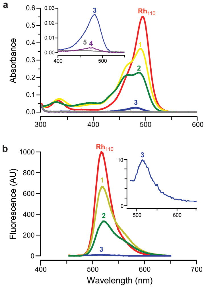
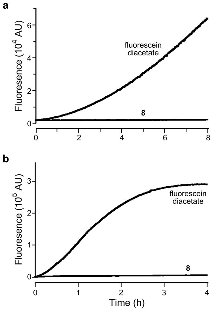
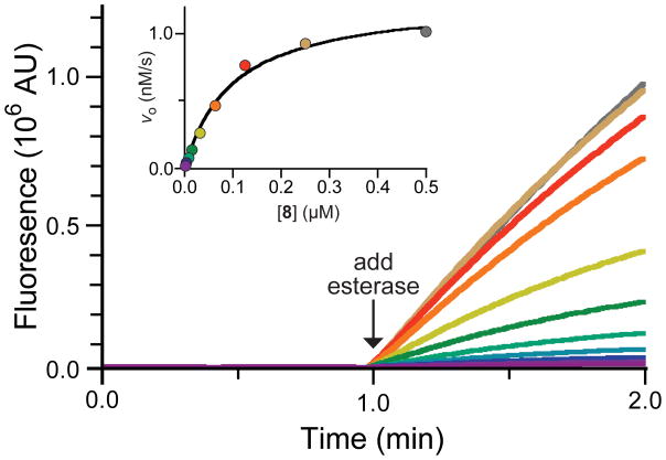
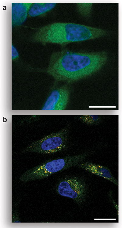
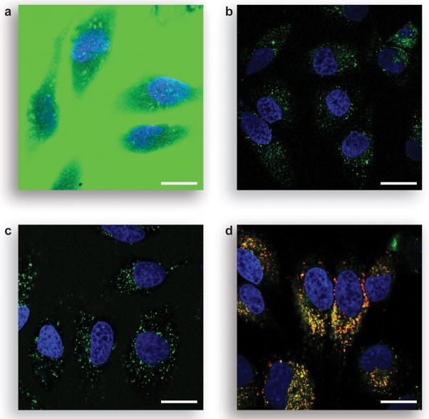
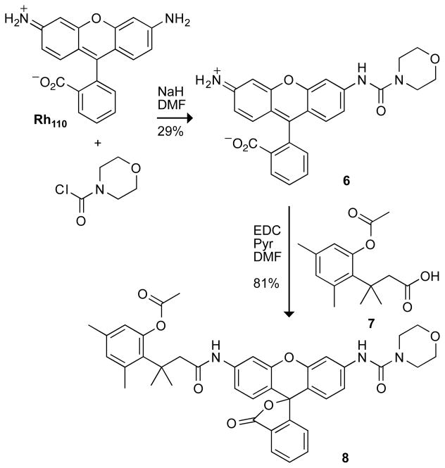
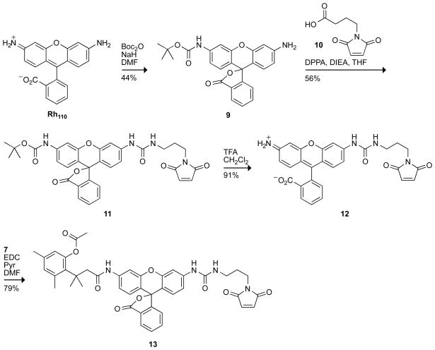
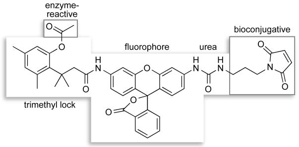
Traditional small-molecule fluorophores are always fluorescent. This attribute can obscure valuable information in biological experiments. Here, we report on a versatile "latent" fluorophore that overcomes this limitation. At the core of the latent fluorophore is a derivative of rhodamine in which one nitrogen is modified as a urea. That modification enables rhodamine to retain half of its fluorescence while facilitating conjugation to a target molecule. The other nitrogen of rhodamine is modified with a "trimethyl lock", which enables fluorescence to be unmasked fully by a single user-designated chemical reaction. An esterase-reactive latent fluorophore was synthesized in high yield and attached covalently to a cationic protein. The resulting conjugate was not fluorescent in the absence of esterases. The enzymatic activity of esterases in endocytic vesicles and the cytosol educed fluorescence, enabling the time-lapse imaging of endocytosis into live human cells and thus providing unprecedented spatiotemporal resolution of this process. The modular design of this "fluorogenic label" enables the facile synthesis of an ensemble of small-molecule probes for the illumination of numerous biochemical and cell biological processes.


Figures

Figure 1
Figure 1
Spectra of Rh110 and its derivatives. a) Absorption spectra of Rh110 and derivatives 15 (7.5 μM). b) Fluorescent emission spectra of Rh110 and 13 (λex = 450 nm, not to scale).
Figure 2
Figure 2
Hammett plot of extinction coefficient (○) and quantum yield (●) versus σp for Rh110, urea 1, and amide 2.
Figure 3
Figure 3
Stability of pro-fluorophore 8 and fluorescein diacetate in aqueous solution. a) Time course for the generation of fluorescence (λex 496 nm, λem 520 nm) of pro-fluorophore 8 (25 nM) and fluorescein diacetate (25 nM) in PBS. b) Time course of the generation of fluorescence (λex 496 nm, λem 520 nm) of pro-fluorophore 8 (25 nM) and fluorescein diacetate (25 nM) in DMEM containing FBS (10% v/v).
Figure 4
Figure 4
Kinetic traces (λex 496 nm, λem 520 nm) and Michaelis–Menten plot (inset) of a serial dilution of pro-fluorophore 8 (0.5 μM → 2 nM) with PLE (2.5 μg/mL).
Figure 5
Figure 5
Unmasking of pro-fluorophore 8 in live human cells. a) Unwashed HeLa cells incubated for 1 h with pro-fluorophore 8 (10 μM) at 37 °C in DMEM and counter-stained with Hoechst 33342. b) Washed HeLa cells incubated for 1 h with pro-fluorophore 8 (10 μM) at 37 °C in DMEM and counter-stained with Hoechst 33342 and LysoTracker Red (5% v/v CO2(g), 100% humidity). Scale bar: 20 μm.
Figure 6
Figure 6
Live-cell imaging experiments with protein conjugates. a) Unwashed HeLa cells incubated for 1 h with Oregon Green–RNase A conjugate (10 μM) at 37 °C in DPBS and counter-stained with Hoechst 33342. b) Washed HeLa cells incubated for 1 h with Oregon Green–RNase A conjugate (10 μM) at 37 °C in DPBS and counter-stained with Hoechst 33342. c) Unwashed HeLa cells incubated for 1 h with fluorogenic label 13–RNase A conjugate (10 μM) at 37 °C in DPBS and counter-stained with Hoechst 33342. d) Washed HeLa cells incubated for 1 h with fluorogenic label 13–RNase A conjugate (10 μM) at 37 °C in DPBS and counter-stained with Hoechst 33342 and LysoTracker Red (5% v/v CO2(g), 100% humidity). Scale bar: 20 μm.
Scheme 1
Scheme 1
Synthetic Route to Pro-Fluorophore 8
Scheme 2
Scheme 2
Synthetic Route to Fluorogenic Label 13
Scheme 3
Scheme 3
Modules in Fluorogenic Label 13

Similar articles

Cited by

References

    1. Haugland RP, Spence MTZ, Johnson ID, Basey A. The Handbook: A Guide to Fluorescent Probes and Labeling Technologies. 10. Molecular Probes; Eugene, OR: 2005.
    1. Chandran SS, Dickson KA, Raines RT. Latent fluorophore based on the trimethyl lock. J Am Chem Soc. 2005;127:1652–1653. - PubMed
    1. Lavis LD, Chao T-Y, Raines RT. Latent blue and red fluorophores based on the trimethyl lock. ChemBioChem. 2006;7:xxx–xxx. - PMC - PubMed
    1. Borchardt RT, Cohen LA. Stereopopulation control. II. Rate enhancement of intramolecular nucleophilic displacement. J Am Chem Soc. 1972;94:9166–9174. - PubMed
    1. Milstein S, Cohen LA. Stereopopulation control. I. Rate enhancement in the lactonizations of o-hydroxyhydrocinnamic acids. J Am Chem Soc. 1972;94:9158–9165. - PubMed

Publication types

MeSH terms