

Isolated lymphoid follicle maturation induces the development of follicular dendritic cells
- PMID: 17163957
- PMCID: PMC2265896
- DOI: 10.1111/j.1365-2567.2006.02508.x
Isolated lymphoid follicle maturation induces the development of follicular dendritic cells
Abstract
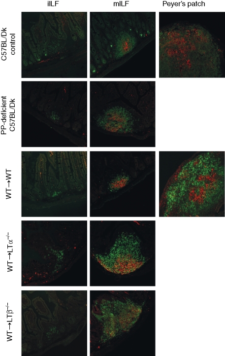
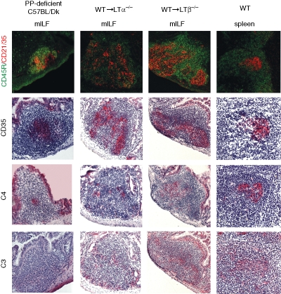
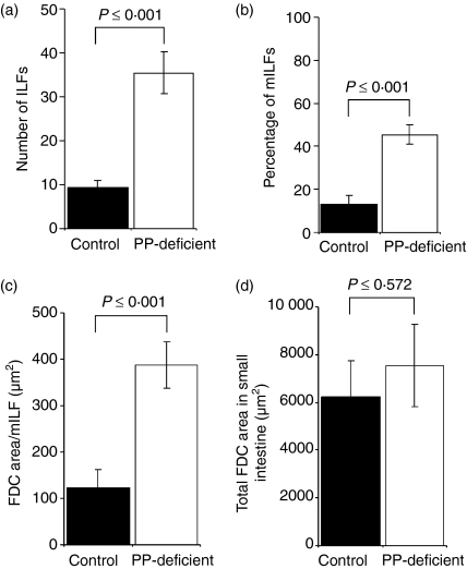
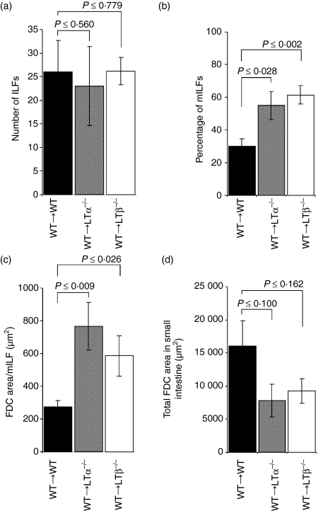
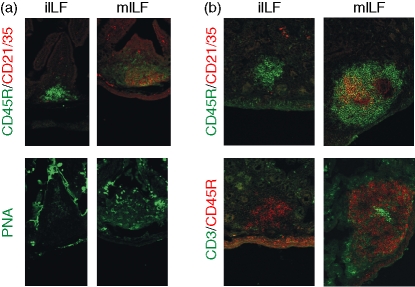
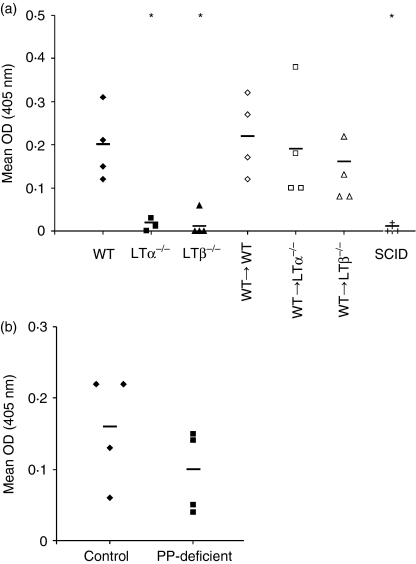
Isolated lymphoid follicles (ILFs) are recently identified lymphoid structures in the small intestine with features similar to Peyer's patches (PPs). Using immunohistochemistry we characterized the composition of ILFs in the small intestines of immunocompetent mice and of mice that lacked PPs as a result of either genetic deficiency of lymphotoxin or temporary in utero lymphotoxin-beta receptor-signalling blockade. We showed that although both immature and mature ILFs were present in the intestines of immunocompetent mice, PP-deficiency induced a significantly greater number of mature ILFs. We found that in addition to B-lymphocyte-containing germinal centres, mature ILFs also possessed large networks of follicular dendritic cells (FDCs). These features were not detected within immature ILFs. Indeed, the presence of FDCs could be used to reliably distinguish ILF maturity. Further analysis revealed that the area occupied by the FDCs within mature ILFs was substantial. The total area occupied by FDCs in all the mature ILFs in mice lacking PPs was equivalent to the total area occupied by FDCs in all the PPs and the few mature ILFs in immunocompetent mice. Based on these data we reasoned that in the absence of PPs, mature ILFs are important inductive sites for intestinal immune responses. Indeed, in mice that lacked PPs, ILF maturation coincided with a restoration of faecal immunoglobulin A levels to values that were comparable to those found in immunocompetent mice. Taken together, these data imply that the induction of germinal centres and FDC networks within mature ILFs in response to PP deficiency provides an important compensatory mechanism.
Figures
Similar articles
-
Isolated lymphoid follicles can function as sites for induction of mucosal immune responses.Ann N Y Acad Sci. 2004 Dec;1029:44-57. doi: 10.1196/annals.1309.006. Ann N Y Acad Sci. 2004. PMID: 15681742
-
Role of the GALT in scrapie agent neuroinvasion from the intestine.J Immunol. 2007 Mar 15;178(6):3757-66. doi: 10.4049/jimmunol.178.6.3757. J Immunol. 2007. PMID: 17339474
-
Adaptive immune responses are dispensable for isolated lymphoid follicle formation: antigen-naive, lymphotoxin-sufficient B lymphocytes drive the formation of mature isolated lymphoid follicles.J Immunol. 2005 May 1;174(9):5720-8. doi: 10.4049/jimmunol.174.9.5720. J Immunol. 2005. PMID: 15843574
-
Induction of intestinal lymphoid tissue: the role of cryptopatches.Ann N Y Acad Sci. 2006 Aug;1072:210-7. doi: 10.1196/annals.1326.015. Ann N Y Acad Sci. 2006. PMID: 17057201 Review.
-
CCR6 and CCL20: partners in intestinal immunity and lymphorganogenesis.Ann N Y Acad Sci. 2006 Aug;1072:52-61. doi: 10.1196/annals.1326.036. Ann N Y Acad Sci. 2006. PMID: 17057190 Review.
Cited by
-
Autophagy at the gut interface: mucosal responses to stress and the consequences for inflammatory bowel diseases.Inflamm Bowel Dis. 2010 Jan;16(1):152-74. doi: 10.1002/ibd.20991. Inflamm Bowel Dis. 2010. PMID: 19575363 Free PMC article. Review.
-
Shigella impairs T lymphocyte dynamics in vivo.Proc Natl Acad Sci U S A. 2013 Mar 19;110(12):4458-63. doi: 10.1073/pnas.1300981110. Epub 2013 Feb 15. Proc Natl Acad Sci U S A. 2013. PMID: 23417297 Free PMC article.
-
Induction of intestinal lymphoid tissue formation by intrinsic and extrinsic signals.Semin Immunopathol. 2009 Jul;31(2):151-69. doi: 10.1007/s00281-009-0163-6. Epub 2009 Jun 9. Semin Immunopathol. 2009. PMID: 19506873 Review.
-
Differential induction of isolated lymphoid follicles in the gut by 18β-glycyrrhetinic acid.PLoS One. 2014 Jul 3;9(7):e100878. doi: 10.1371/journal.pone.0100878. eCollection 2014. PLoS One. 2014. PMID: 24992099 Free PMC article.
-
The Gut-Associated Lymphoid Tissues in the Small Intestine, Not the Large Intestine, Play a Major Role in Oral Prion Disease Pathogenesis.J Virol. 2015 Sep;89(18):9532-47. doi: 10.1128/JVI.01544-15. Epub 2015 Jul 8. J Virol. 2015. PMID: 26157121 Free PMC article.
References
-
- Macpherson AJ, Hunzinker L, McKoy K, Lamarre A. IgA responses in the intestinal mucosa against pathogenic and non-pathogenic microorganisms. Microbes Infect. 2001;3:1021–35. - PubMed
-
- Shikina T, Hiroi T, Iwatani K, et al. IgA class switch occurs in the organized nasopharynx- and gut-associated lymphoid tissue, but not in the diffuse lamina propria or airways and gut. J Immunol. 2004;172:6259–64. - PubMed
-
- Yamamoto M, Rennert P, McGhee JR, et al. Alternate mucosal immune system: organized Peyer's patches are not required for IgA responses in the gastrointestinal tract. J Immunol. 2000;164:5184–91. - PubMed
-
- Hamada H, Hiroi T, Nishiyama Y, et al. Identification of multiple isolated lymphoid follicles on the antimesenteric wall of the mouse small intestine. J Immunol. 2002;168:57–64. - PubMed
-
- Kweon MN, Yamamoto M, Rennert PD, et al. Prenatal blockage of lymphotoxin β receptor and TNF receptor p55 signaling cascade resulted in the acceleration of tissue genesis for isolated lymphoid follicles in the large intestine. J Immunol. 2005;174:4365–72. - PubMed
Publication types
MeSH terms
Substances
Grants and funding
- BBS/E/A/00001660/BB_/Biotechnology and Biological Sciences Research Council/United Kingdom
- G9721848/MRC_/Medical Research Council/United Kingdom
- BBS/E/A/00001659/BB_/Biotechnology and Biological Sciences Research Council/United Kingdom
- BB/D00831X/2/BB_/Biotechnology and Biological Sciences Research Council/United Kingdom
- BBS/E/I/00000989/BB_/Biotechnology and Biological Sciences Research Council/United Kingdom
LinkOut - more resources
Full Text Sources
Molecular Biology Databases
Miscellaneous
