. 2007 Jan;63(2):363-78.
doi: 10.1111/j.1365-2958.2006.05518.x. Epub 2006 Dec 5.

Characterization of the Thermotoga maritima chemotaxis methylation system that lacks pentapeptide-dependent methyltransferase CheR:MCP tethering

Affiliations

Characterization of the Thermotoga maritima chemotaxis methylation system that lacks pentapeptide-dependent methyltransferase CheR:MCP tethering

Eduardo Perez et al. Mol Microbiol. 2007 Jan.

Abstract

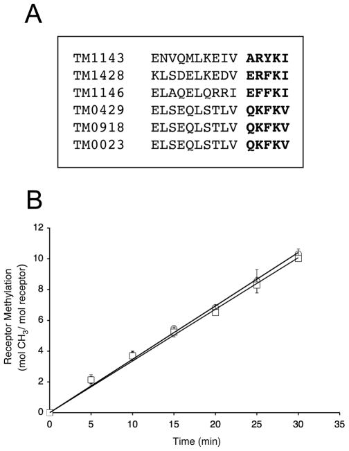
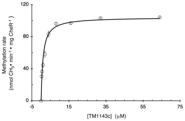
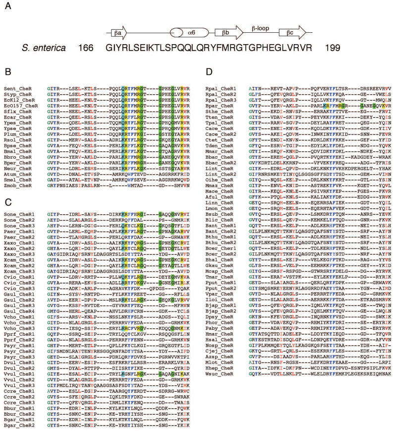
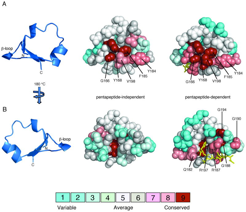
Sensory adaptation in bacterial chemotaxis is mediated by covalent modifications of specific glutamate and glutamine residues within the cytoplasmic domains of methyl-accepting chemotaxis proteins (MCPs). In Escherichia coli and Salmonella enterica, efficient methylation of MCPs depends on the localization of methyltransferase CheR to MCP clusters through an interaction between the CheR beta-subdomain and a pentapeptide sequence (NWETF or NWESF) at the C-terminus of the MCP. In vitro methylation analyses utilizing S. enterica and Thermotoga maritima CheR proteins and MCPs indicate that MCP methylation in T. maritima occurs independently of a pentapeptide-binding motif. Kinetic and binding measurements demonstrate that despite efficient methylation, the interaction between T. maritima CheR and T. maritima MCPs is of relatively low affinity. Comparative protein sequence analyses of CheR beta-subdomains from organisms having MCPs that contain and/or lack pentapeptide-binding motifs identified key similarities and differences in residue conservation, suggesting the existence of two distinct classes of CheR proteins: pentapeptide-dependent and pentapeptide-independent methyltransferases. Analysis of MCP C-terminal ends showed that only approximately 10% of MCPs contain a putative C-terminal binding motif, the majority of which are restricted to the different proteobacteria classes (alpha, beta, gamma, delta). These findings suggest that tethering of CheR to MCPs is a relatively recent event in evolution and that the pentapeptide-independent methylation system is more common than the well-characterized pentapeptide-dependent methylation system.


Figures

FIG. 1
FIG. 1. Analysis of T. maritima receptor C-terminal ends and their role in methylation
A. Sequence alignment of T. maritima transmembrane receptor C-terminal ends. A multiple sequence alignment was generated with ClustalW (Thompson et al., 1994) using the highly conserved domain (HCD) core (Zhulin, 2001) as the anchor for alignment. The last fifteen residues of each of receptor are shown, with the last five residues in bold. B. Methylation of TM1428 and TM1428Δ5. Methyltransferase activity was determined as described in Experimental Procedures using TM1428 (O) and TM1428Δ5 (□) as substrates. Assays were performed with 1.95 and 3.90 pmol of T. maritima CheR and the data shown are the average values from three independent experiments with standard errors.
FIG. 2
FIG. 2. Kinetics of methylation of TM1143c catalyzed by T. maritima CheR
Methyltransferase activity was determined as described in Experimental Procedures by incubating different concentrations of TM1143c (0 to 64 μM) with 0.20 μM methyltransferase CheR for 25 min at 30°C. Km and Kcat values of 1.14 ± 0.20 μM and 3.60 ± 0.40 mol CH3·min-1·mg CheR-1 were estimated by fitting data to the Michaelis-Menton equation using SigmaPlot 8.0. The data shown are the average values of initial velocities from three experiments with standard errors and the fitted curve used for estimation of Km and Kcat.
FIG. 3
FIG. 3. Multiple sequence alignments of CheR β-subdomains
A. Secondary structure diagram of the S. enterica CheR β-subdomain derived from crystal structures (Djordjevic and Stock, 1997, Djordjevic, 1998 #1644) with the amino acid sequence and residue numbers indicated. A multiple sequence alignment for all β-subdomain sequences was generated with ClustalW (Thompson et al., 1994) and the β-subdomain sequences were categorized as described in the text. B. Group I: β-subdomains from organisms containing only 1 CheR protein and ≥1 MCP with a putative pentapeptide sequence. C. Group II: β-subdomains from organisms containing ≥2 CheR proteins and ≥1 MCP with a putative pentapeptide sequence. D. Group III: β-subdomains from organisms containing ≥1 CheR and only MCPs that lacked putative pentapeptide sequences. Conserved amino acid residues in all β-subdomain sequences (≥85%) are colored: small (A, G), green; hydrophobic (L, V, I, M, F, Y, W, H), red; and aromatic (Y, F, W, H), blue; with the most common residue at each position in bold. Amino acid residues that are conserved and that are proposed to be important for CheR-pentapeptide interactions are highlighted: small (G), green; positively charged (R, K), yellow; and side chain amine/amide containing residues (Q, K, R), cyan. The organism abbreviation for each CheR homolog and the corresponding TIGR identification number are: Aful, Archaeoglobus fulgidus (AF1037); Atum, Agrobacterium tumefaciens (NT02AT0609); Azsp, Azoarcus sp. EbN1 (NT01AE1293); Bant, Bacillus anthracis Ames(BA1665, BA0995); Bbac, Bdellovibrio bacteriovorus (NT02BB3177, NT02BB2588); Bbro, Bordetella bronchiseptica (NT01BB2503); Bbur, Borrelia burgdorferi (BB0040, BB0414); Bcer, Bacillus cereus (NT01BC1535, NT01BC0909); Bgar, Borrelia garinii (NT01BG0039, NT01BG0418); Bhal, Bacillus halodurans (NT01BH1876); Bjap, Bradyrhizobium japonicum (NT01BJ0473, NT01BJ2738); Blic, Bacillus licheniformis (NT03BL2556); Bmal, Burkholderia mallei (BMA2856); Bpar, Bordetella parapertussis (NT02BP1517); Bper, Bordetella pertussis (NT03BP1055); Bpse, Burkholderia pseudomallei (ntbp3311); Bsub, Bacillus subtilis (NT01BS2884); Bthu, Bacillus thuringiensis (NT02BT1675, NT02BT1053); Cace, Clostridium acetobutylicum (NT01CA2443, NT01CA0131); Ccre, Caulobacter crescentus (CC0435, CC0598, CC3472); Cjej, Campylobacter jejuni (NT01CJ0929); Ctet, Clostridium tetani (NT02CT1848); Cvio, Chromobacterium violaceum (NT01CV2459, NT01CV3397, NT01CV3658); Dpsy, Desulfotalea psychrophila (NT01DP3085); Dvul, Desulfovibrio vulgaris (DVU1595, DVU2076); Ecar, Erwinia carotovora (NT06EC1756); EcK12, Escherichia coli K12 (NT01EC2290); Ec0157, Escherichia coli O157:H7 (NT02EC2821); Gkau, Geobacter kaustophilus (NT01GK2440); Gsul, Geobacter sulfurreducens (GSU0295, GSU1143, GSU2215, GSU3195); Hhep, Helicobacter hepaticus (NT02HP0476); Hmar, Haloarcula marismortui (NT01HMA2142); Hsal, Halobacterium salinarum (NT01HS0757); Iloi, Idiomarina loihiensis (NT01IL1191); Linn, Listeria innocua (NT01LI0715); Lint, Leptospira interrogans (NT02LI2313, NT02LI1981); Lmon, Listeria monocytogenes (NT01LM0742); Mace, Methanosarcina acetivorans (NT02MA3813); Mcap, Methylococcus capsulatus (MCA0829); Mmar, Methanococcus maripaludis (NT04MM0987); Mlot, Mesorhizobium loti (NT02MLB0021); Mmaz, Methanosarcina mazei (NT01MM1835); Neur, Nitrosomonas europaea (NT01NE2059); Nosp, Nostoc sp. PCC7120 (NT01NS2346); Oihe, Oceanobacillus iheyensis (NT01OI1946); Paby, Pyrococcus abyssi (NT01PA1746); Paer, Pseudomonas aeruginosa (NT03PA0199, NT03PA3856); Phor, Pyrococcus horikoshii (NT01PH0504); Plum, Photorhabdus luminescens (NT01PL2011); Pprf, Photobacterium profundum (NT01PP0812, NT01PP0945); Pput, Pseudomonas putida (PP4392, PP3760); Psyr, Pseudomonas syringae (PSPPH0802, PSPH2602, PSPPH3412); Rpal, Rhodopseudomonas palustris (NT02RP0142, NT02RP1682, NT02RP1731); Rsol, Ralstonia solanacearum (NT01RSA1413); Sent, Salmonella enterica serovar Typhimurium CT18 (NT03ST2156); Sflx, Shigella flexneri (NT02SF2178); Smel, Sinorhizobium meliloti (NT01SM0883); Sone, Shewanella oneidensis (SO2124, SO3251, SO2325); Sthe, Symbiobacterium thermophilum (NT07ST1815); Styp, Salmonella Typhimurium LT2 (NT05SE0993); Tden, Treponema denticola (TDE0647); Tmar, Thermotoga maritima (TM0464); Tpal, Treponema pallidum (TP0630); Tten, Thermoanaerobacter tengcongens (NT01TT1507);Vcho; Vibrio cholerae (VC1399, VC2201, VCA1091); Vpar, Vibrio parahaemolyticus (NT01VP0730); Vvul, Vibrio vulnificus (NT01VVA1160, NT01VV0231, NT01VVA0376); Wsuc, Wolinella succinogenes (NT01WS1194); Xaxo, Xanthomonas axonopodis (NT01XA2561, NT01XA3795, NT01XA1741); Xcam, Xanthomonas campestris (NT01XC2547, NT01XC3635, NT01XC1630); Ypes, Yersinia pestis (NT02YP2177); Ypse, Yersinia pseudotuberculosis (NT05YP2660); Zmob, Zymomonas mobilis (NT01ZM0078).
FIG. 4
FIG. 4
Amino acid conservation on the surfaces of pentapeptide-independent and pentapeptide-dependent β-subdomains. Multiple sequence alignments of pentapeptide-independent and pentapeptide-dependent CheR β-subdomains, classified in Figure 3, were generated using ClustalW (Thompson et al., 1994). Amino acid conservation scores for each residue were calculated using the Consurf maximum likelihood method (Glaser et al., 2003) (http://consurf.tau.ac.il) and were mapped onto space-filling models of the CheR β-subdomain (PDB accession code 1BC5) (Djordjevic and Stock, 1998) using Pymol (Delano, 2002). The coloring scheme for conservation ranges from cyan (variable) to red (conserved) and the pentapeptide (gold) is shown in stick representation. A. β-subdomain interior face. The ribbon diagram of the β-subdomain (left) indicates the orientation of the space-filling models, viewed toward the surface that packs against the catalytic domain of CheR. Residues that are highly conserved in both pentapeptide-independent (center) and pentapeptide-dependent (right) β-subdomains are labeled. B. β-subdomain exterior face. The orientation corresponds to a 180° rotation around a vertical axis relative to the view in A. Residues that are highly conserved only in pentapeptide-dependent β-subdomains (right) are labeled.
FIG. 5
FIG. 5. Conservation of residues within the β-subdomain of CheR
The CheR-pentapeptide structure (left) is displayed as a space-filling model (green), except for the β-subdomain (blue) and pentapeptide (gold), which are shown as a ribbon diagram and in stick representation, respectively. An enlarged view of the β-subdomain (right) shows conserved residues in stick representation. Universally conserved β-subdomain residues are shown in orange and residues that are conserved exclusively in pentapeptide-dependent β-subdomains are shown in red. Images were generated using Pymol (Delano, 2002).

Similar articles

Cited by

References

    1. Alley MRK, Maddock JR, Shapiro L. Polar localization of a bacterial chemoreceptor. Genes and Development. 1992;6:825–836. - PubMed
    1. Anand GS, Stock AM. Kinetic basis for the stimulatory effect of phosphorylation on the methylesterase activity of CheB. Biochemistry. 2002;41:6752–6760. - PubMed
    1. Armitage JP, Schmitt R. Bacterial chemotaxis: Rhodobacter sphaeroides and Sinorhizobium meliloti-variations on a theme? Microbiology. 1997;143 (Pt 12):3671–3682. - PubMed
    1. Astling DP, Lee JY, Zusman DR. Differential effects of chemoreceptor methylation-domain mutations on swarming and development in the social bacterium Myxococcus xanthus. Mol Microbiol. 2006;59:45–55. - PubMed
    1. Banno S, Shiomi D, Homma M, Kawagishi I. Targeting of the chemotaxis methylesterase/deamidase CheB to the polar receptor-kinase cluster in an Escherichia coli cell. Mol Microbiol. 2004;53:1051–1063. - PubMed

Publication types

MeSH terms