

Rb induces a proliferative arrest and curtails Brn-2 expression in retinoblastoma cells
- PMID: 17163992
- PMCID: PMC1764425
- DOI: 10.1186/1476-4598-5-72
Rb induces a proliferative arrest and curtails Brn-2 expression in retinoblastoma cells
Abstract
Background: Retinoblastoma is caused by loss of the Rb protein in early retinal cells. Although numerous Rb functions have been identified, Rb effects that specifically relate to the suppression of retinoblastoma have not been defined.
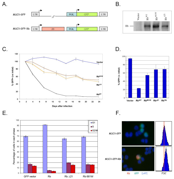
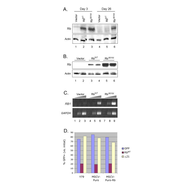
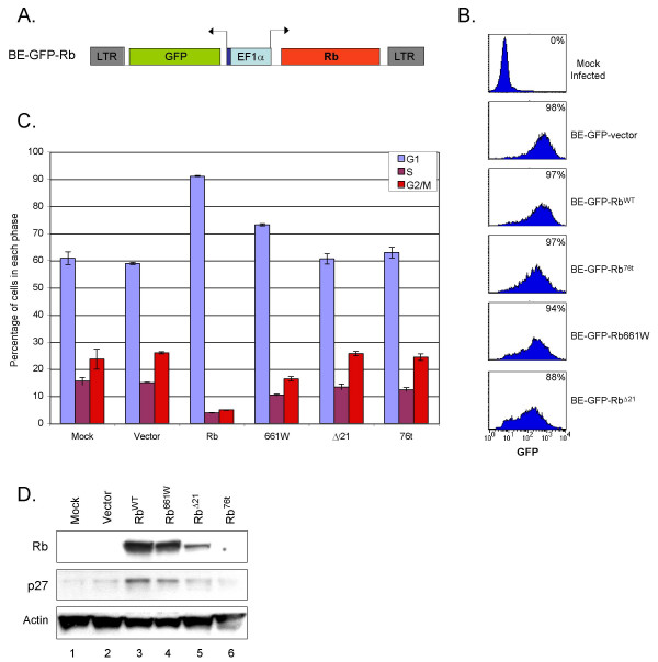
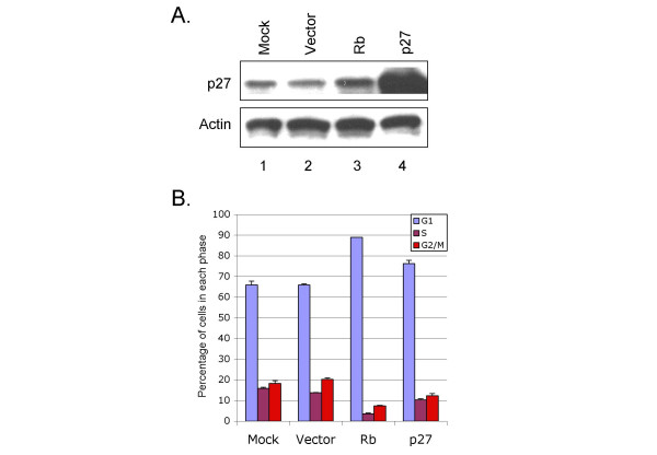
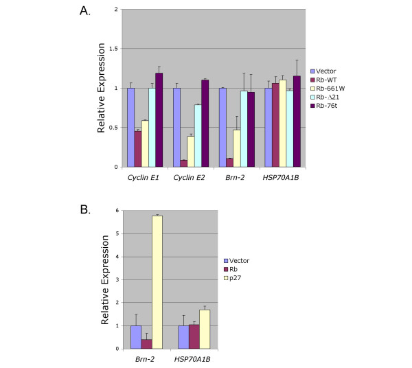
Results: In this study, we examined the effects of restoring Rb to Y79 retinoblastoma cells, using novel retroviral and lentiviral vectors that co-express green fluorescent protein (GFP). The lentiviral vector permitted transduction with sufficient efficiency to perform biochemical analyses. Wild type Rb (RbWT) and to a lesser extent the low penetrance mutant Rb661W induced a G0/G1 arrest associated with induction of p27KIP1 and repression of cyclin E1 and cyclin E2. Microarray analyses revealed that in addition to down-regulating E2F-responsive genes, Rb repressed expression of Brn-2 (POU3F2), which is implicated as an important transcriptional regulator in retinal progenitor cells and other neuroendocrine cell types. The repression of Brn-2 was a specific Rb effect, as ectopic p27 induced a G0/G1 block, but enhanced, rather than repressed, Brn-2 expression.
Conclusion: In addition to Rb effects that occur in many cell types, Rb regulates a gene that selectively governs the behavior of late retinal progenitors and related cells.
Figures
Similar articles
-
Lentiviral vector-mediated PAX6 overexpression promotes growth and inhibits apoptosis of human retinoblastoma cells.Invest Ophthalmol Vis Sci. 2011 Oct 28;52(11):8393-400. doi: 10.1167/iovs.11-8139. Invest Ophthalmol Vis Sci. 2011. PMID: 21948554
-
Co-deleting Pten with Rb in retinal progenitor cells in mice results in fully penetrant bilateral retinoblastomas.Mol Cancer. 2015 Apr 24;14:93. doi: 10.1186/s12943-015-0360-y. Mol Cancer. 2015. PMID: 25907958 Free PMC article.
-
Inhibition of Hsp90 function by ansamycins causes retinoblastoma gene product-dependent G1 arrest.Cancer Res. 2000 Jul 15;60(14):3940-6. Cancer Res. 2000. PMID: 10919672
-
Using kinetic studies to uncover new Rb functions in inhibiting cell cycle progression.Cell Cycle. 2005 Mar;4(3):373-5. doi: 10.4161/cc.4.3.1535. Epub 2005 Mar 13. Cell Cycle. 2005. PMID: 15701969 Review.
-
Targeted pharmacologic inhibition of S-phase kinase-associated protein 2 (SKP2) mediated cell cycle regulation in lung and other RB-Related cancers: A brief review of current status and future prospects.Adv Biol Regul. 2023 May;88:100964. doi: 10.1016/j.jbior.2023.100964. Epub 2023 Mar 14. Adv Biol Regul. 2023. PMID: 37004354 Review.
Cited by
-
Rb suppresses human cone-precursor-derived retinoblastoma tumours.Nature. 2014 Oct 16;514(7522):385-8. doi: 10.1038/nature13813. Epub 2014 Sep 24. Nature. 2014. PMID: 25252974 Free PMC article.
-
Structural and functional analysis of cancer-associated missense variants in the retinoblastoma protein pocket domain.J Biol Chem. 2025 Mar;301(3):108284. doi: 10.1016/j.jbc.2025.108284. Epub 2025 Feb 10. J Biol Chem. 2025. PMID: 39938803 Free PMC article.
-
Retinoblastoma has properties of a cone precursor tumor and depends upon cone-specific MDM2 signaling.Cell. 2009 Jun 12;137(6):1018-31. doi: 10.1016/j.cell.2009.03.051. Cell. 2009. PMID: 19524506 Free PMC article.
-
Reciprocal Induction of MDM2 and MYCN in Neural and Neuroendocrine Cancers.Front Oncol. 2020 Dec 23;10:563156. doi: 10.3389/fonc.2020.563156. eCollection 2020. Front Oncol. 2020. PMID: 33425720 Free PMC article.
-
Resolving the current controversy of use and reuse of housekeeping proteins in ageing research: Focus on saving people's tax dollars.Ageing Res Rev. 2024 Sep;100:102437. doi: 10.1016/j.arr.2024.102437. Epub 2024 Jul 25. Ageing Res Rev. 2024. PMID: 39067773 Review.
References
Publication types
MeSH terms
Substances
LinkOut - more resources
Full Text Sources
