Comparative Study
. 2006 Dec 19;103(51):19478-83.
doi: 10.1073/pnas.0609329103. Epub 2006 Dec 12.

The small-molecule VEGF receptor inhibitor pazopanib (GW786034B) targets both tumor and endothelial cells in multiple myeloma

Affiliations
Comparative Study

The small-molecule VEGF receptor inhibitor pazopanib (GW786034B) targets both tumor and endothelial cells in multiple myeloma

Klaus Podar et al. Proc Natl Acad Sci U S A. .

Abstract

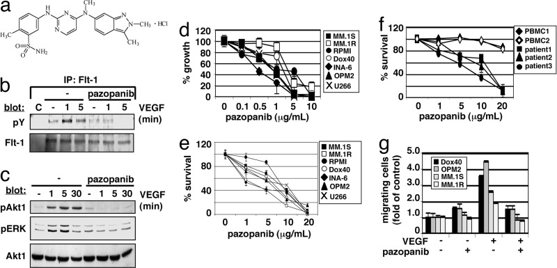
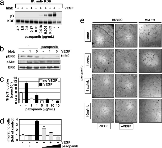
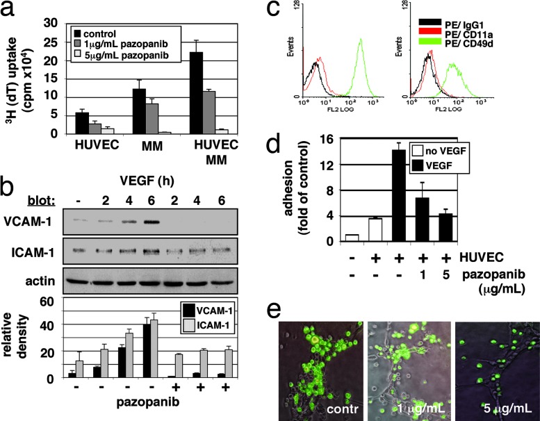
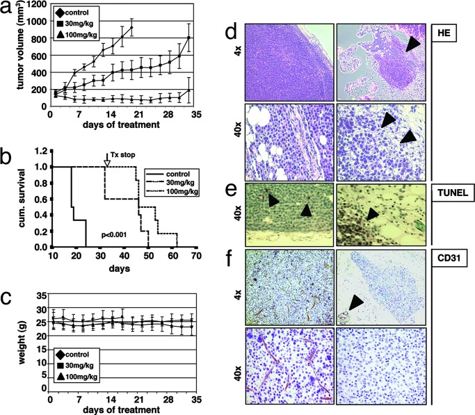
A critical role for vascular endothelial factor (VEGF) has been demonstrated in multiple myeloma (MM) pathogenesis. Here, we characterized the effect of the small-molecule VEGF receptor inhibitor pazopanib on MM cells in the bone marrow milieu. Pazopanib inhibits VEGF-triggered signaling pathways in both tumor and endothelial cells, thereby blocking in vitro MM cell growth, survival, and migration, and inhibits VEGF-induced up-regulation of adhesion molecules on both endothelial and tumor cells, thereby abrogating endothelial cell-MM cell binding and associated cell proliferation. We show that pazopanib is the first-in-class VEGF receptor inhibitor to inhibit in vivo tumor cell growth associated with increased MM cell apoptosis, decreased angiogenesis, and prolonged survival in a mouse xenograft model of human MM. Low-dose pazopanib demonstrates synergistic cytotoxicity with conventional (melphalan) and novel (bortezomib and immunomodulatory drugs) therapies. Finally, gene expression and signaling network analysis show transcriptional changes of several cancer-related genes, in particular c-Myc. Using siRNA, we confirm the role of c-Myc in VEGF production and secretion, as well as angiogenesis. These preclinical studies provide the rationale for clinical evaluation of pazopanib, alone and in combination with conventional and novel therapies, to increase efficacy, overcome drug resistance, reduce toxicity, and improve patient outcome in MM.


Conflict of interest statement

Conflict of interest statement: R.K. and L.N.P. are employees of GlaxoSmithKline, Research Triangle Park, NC.

Figures

Fig. 1.
Fig. 1.
Pazopanib inhibits MM cell growth, survival, and migration via VEGF-triggered Flt-1 phosphorylation and activation of downstream signaling molecules. (a) Chemical structure of pazopanib (GW786034B). (b) Pazopanib inhibits VEGF-induced Flt-1 autophosphorylation. (c) Pazopanib inhibits VEGF-induced phosphorylation of Akt1 and ERK. (d) Pazopanib induces inhibition of MM cell growth. Values represent mean ± SD 3H[dT] uptake of triplicate cultures. (e and f) Pazopanib triggers MM cell cytotoxicity in MM cell lines (e) and CD138 plus patient cells, but not in normal PBMCs (f). MTT cleavage was measured during the last 4 h of a 48-h culture. Values represent the mean ± SD of quadruplicate cultures. (g) Pazopanib inhibits VEGF-triggered MM cell migration. Growth factor-deprived MM.1S, MM.1R, RPMI-Dox40 (Dox40), and OPM2 cells were pretreated with pazopanib or left untreated, plated on a fibronectin-coated polycarbonate membrane (8-μm pore size) in a modified Boyden chamber, and exposed to 10 ng/ml VEGF for 4 to 6 h.
Fig. 2.
Fig. 2.
Effects of pazopanib on endothelial cells. (a and b) Pazopanib inhibits VEGF-induced phosphorylation of KDR (a) and downstream activation of signaling molecules (b). (c) Pazopanib inhibits VEGF-triggered HUVEC proliferation. Values represent the mean ± SD 3H[dT] uptake of triplicate cultures. (d) Pazopanib inhibits VEGF-triggered HUVEC migration. Growth factor-deprived HUVECs were pretreated with pazopanib (1, 5, and 10 μg/ml) or were left untreated. Cells then were placed on a polycarbonate membrane (8-μm pore size) in a modified Boyden chamber and were exposed for 6–10 h to 10 ng/ml VEGF. At the end of treatment, cells on the lower part of the membrane were counted by using a Coulter counter ZBII. Data represent mean ± SD for duplicate samples and are representative of three independent experiments. (e) Pazopanib inhibits VEGF-triggered endothelial tubule formation. HUVEC suspensions (with or without VEGF) or endothelial cells isolated from BM aspirates (MM EC) were premixed with different concentrations of pazopanib and added on top of the ECMatrix. Tube formation was assessed by using an inverted light microscope at ×4 and ×10 magnification. Photographs are representative of each group and three independent experiments.
Fig. 3.
Fig. 3.
Effect of pazopanib on endothelial-MM coculture systems. (a) Pazopanib inhibits proliferation of MM cells adherent to HUVECs. Data shown are mean ± SD of experiments performed in triplicate. (b) Pazopanib induces inhibition of VEGF-mediated up-regulation of surface adhesion proteins VCAM-1 and ICAM-1. Densitometry was used to quantitate expression data of three separate experiments. (c) Pazopanib triggers down-regulation of MM adhesion proteins. Control (c Left) MM.1S cells treated with pazopanib (5 μg/ml) (c Right) were examined for LFA-1 (CD11a/CD18) and VLA-4 (CD49d/CD29) expression by using immunofluorescence flow cytometric analysis. (d) Pazopanib induces dose-dependent inhibition of VEGF-induced MM cell adhesion on HUVECs. (e) Pazopanib decreases adhesion of MM cells in the branching points of endothelial tubule. HUVECs were cultured onto matrigel in EGM-2 medium supplemented with 2% FBS. After formation of tubules, MM/EGFP cells were added with or without pazopanib (1 and 5 μg/ml) for 6–10 h. After removing nonadherent cells, photographs of HUVECs were overlaid with photographs of MM/EGFP cells. Representative photographs (×10 magnification) of each group are shown.
Fig. 4.
Fig. 4.
Pazopanib prolongs survival in a xenograft mouse model by induction of tumor cell apoptosis and inhibition of tumor angiogenesis. Beige-nude Xid mice were inoculated s.c. with 3 × 10⁷ MM.1S MM cells. Daily treatment by oral gavage (vehicle alone, 30 mg/kg, and 100 mg/kg) was started when tumors were measurable. (a) Tumor burden was measured every other day by using a caliper [calculated volume = (4π / 3) × (width / 2)2 × (length / 2)]. Tumor volume is presented as means ± SE. (b) Survival was evaluated by using Kaplan–Meier curves and log-rank analysis. (c) Body weight was evaluated 3 times a week. (df) Representative microscopic images of tumor sections are shown stained with hematoxylin/eosin (HE) (d), TUNEL (e), and anti-CD31 (f).

References

    1. Folkman J. N Engl J Med. 1971;285:1182–1186. - PubMed
    1. Folkman J. Semin Oncol. 2001;28:536–542. - PubMed
    1. Podar K, Anderson KC. Blood. 2005;105:1383–1395. - PubMed
    1. Ferrara N, Gerber HP, LeCouter J. Nat Med. 2003;9:669–676. - PubMed
    1. Kyle RA, Rajkumar SV. N Engl J Med. 2004;351:1860–1873. - PubMed

Publication types

MeSH terms