. 2007 Jan;134(1):167-76.
doi: 10.1242/dev.02701.

Dusp6 (Mkp3) is a negative feedback regulator of FGF-stimulated ERK signaling during mouse development

Affiliations

Dusp6 (Mkp3) is a negative feedback regulator of FGF-stimulated ERK signaling during mouse development

Chaoying Li et al. Development. 2007 Jan.

Abstract

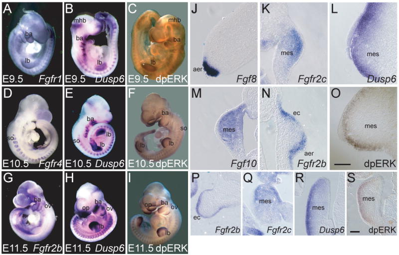
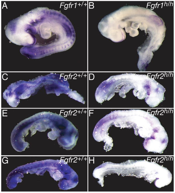
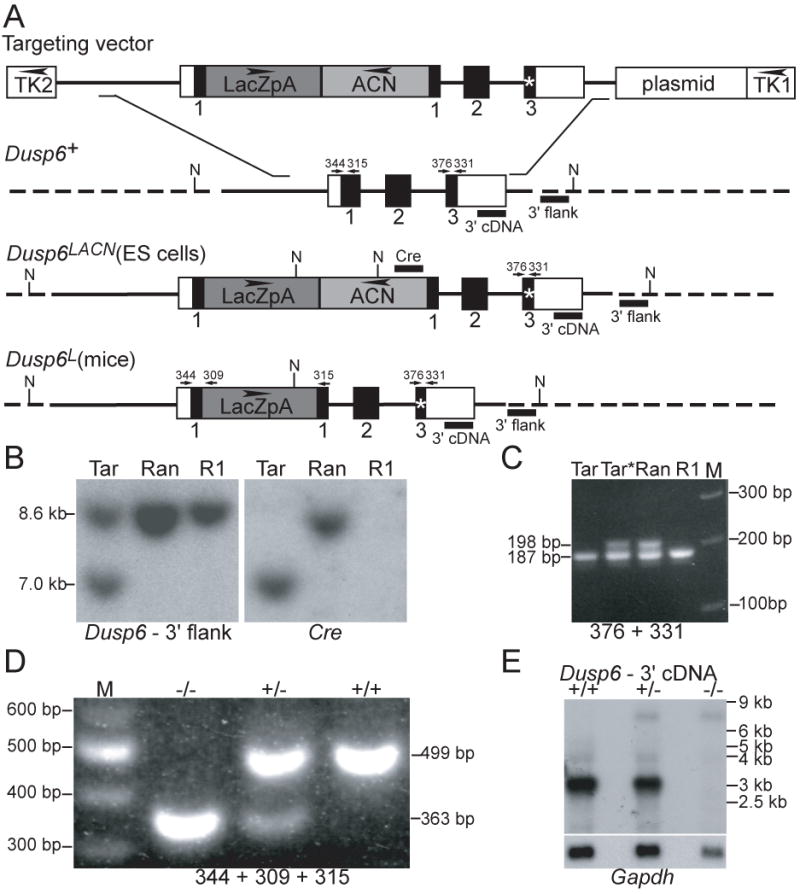
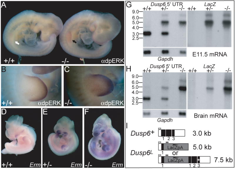
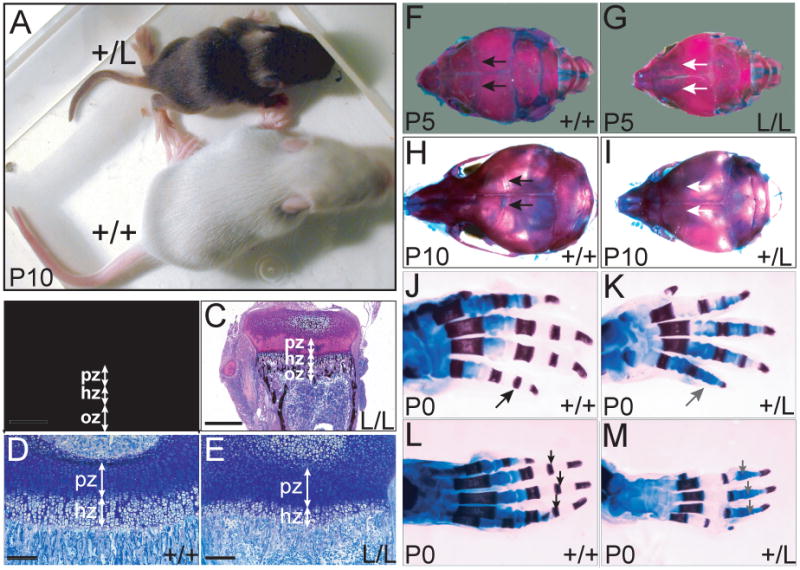
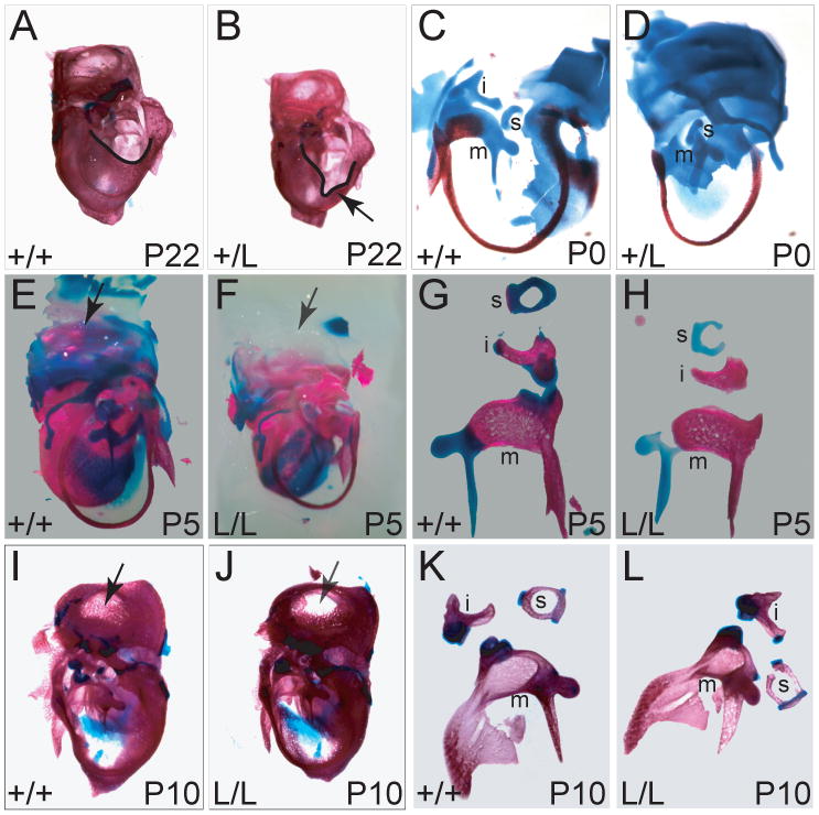
Mitogen-activated protein kinase (MAPK) pathways are major mediators of extracellular signals that are transduced to the nucleus. MAPK signaling is attenuated at several levels, and one class of dual-specificity phosphatases, the MAPK phosphatases (MKPs), inhibit MAPK signaling by dephosphorylating activated MAPKs. Several of the MKPs are themselves induced by the signaling pathways they regulate, forming negative feedback loops that attenuate the signals. We show here that in mouse embryos, Fibroblast growth factor receptors (FGFRs) are required for transcription of Dusp6, which encodes MKP3, an extracellular signal-regulated kinase (ERK)-specific MKP. Targeted inactivation of Dusp6 increases levels of phosphorylated ERK, as well as the pERK target, Erm, and transcripts initiated from the Dusp6 promoter itself. Finally, the Dusp6 mutant allele causes variably penetrant, dominant postnatal lethality, skeletal dwarfism, coronal craniosynostosis and hearing loss; phenotypes that are also characteristic of mutations that activate FGFRs inappropriately. Taken together, these results show that DUSP6 serves in vivo as a negative feedback regulator of FGFR signaling and suggest that mutations in DUSP6 or related genes are candidates for causing or modifying unexplained cases of FGFR-like syndromes.


Figures

Figure 1
Figure 1
Dusp6 expression correlates with particular Fgfrs and with most areas of dpERK expression. (A,D,G) Whole mount RNA in situ hybridization (WmISH) of mouse embryos with the Fgfr probes indicated at the bottom right of each panel. (B,E,H) WmISH of embryos with a Dusp6 probe. (C,F,I) Whole mount immunostaining of embryos with antibody directed against diphosphorylated ERK (dpERK). Embryo age is indicated at the bottom left of each panel. (J-R) Transverse sections of E10-E10.5 WmISH embryos illustrating potential FGF signaling pathways in the limb bud (J-O) and branchial arches (P-R). Probes are noted at the bottom left of each panel. Abbreviations: ba, branchial arches; lb, limb bud; mhb, mid-hindbrain junction; so, somites; ov, otic vesicle; op, olfactory pit; aer, apical ectodermal ridge; ec, ectoderm; mes, mesenchyme.
Figure 2
Figure 2
Dusp6 expression depends on signaling through FGF receptors. WmISH of wild type and homozygous Fgfr hypomorphic (h) mutant littermates with a Dusp6 probe (A,B) Fgfr1α, E9.5; (C-H) Fgfr2ΔIgIII, E8.5. Genotype of each embryo is indicated in the upper left of each panel.
Figure 3
Figure 3
Gene targeting at the Dusp6 locus. (A) Structure of the linearized Dusp6 targeting vector and depiction of the wild type Dusp6 allele (Dusp6+), the correctly targeted mutant allele in ES cells (Dusp6LACN) and the targeted allele found in mice following expression of CRE in germline-transmitting chimeras (Dusp6L). Mouse genomic Dusp6 DNA is depicted with solid thick lines; dotted lines indicate Dusp6 genomic DNA not present in the targeting vector; open boxes indicate untranslated regions; solid boxes indicate protein coding regions. The LacZ gene (nls-lacZpA) is shown as a dark grey box; the Cre/Neo “suicide cassette” (ACN) as a light grey box; the stop codon in the DUSP6 frame in exon 3 as an asterisk. Flanking thymidine kinase genes (TK1 and TK2, transcriptional orientation indicated by arrows) and the plasmid backbone are depicted as open boxes. Recognition sites for Nde I are indicated by “N”; probes used for Southern analysis by black bars. Numbered arrows indicate the identity, position and directionality of primers used in PCR assays. (B) Southern blot hybridization assay demonstrating correct targeting of Dusp6 in ES cells. Nde I-digested DNA from the R1 ES cell line (R1), a cell line with a random insertion of the targeting vector (Ran) and a targeted cell line (Tar) was probed sequentially with 3′ and Cre probes. Correctly targeted cell lines had a novel 7.0 kb fragment that hybridized with both probes. (C) PCR assay used to detect the stop-codon-containing insertion in exon 3. DNAs isolated from correctly targeted ES cells (Tar), a control random integrant (Ran) and wild type cells (R1) were PCR-amplified using primers 376 and 331. Targeted cell lines (TAR*) that produced both the wild type (187 bp) and the insertion amplicon (198 bp) were selected for germline transmission. (D) PCR assay used to genotype offspring of Dusp6+/L intercrosses. Tail DNAs were PCR-amplified with primers 344, 309 and 315. The mutant allele yielded a 363 bp band and the wild type allele yielded a 499 bp band. (E) Northern blot hybridization of mRNA isolated from E11.5 embryos of the indicated genotypes was probed with a fragment of Dusp6 3′ UTR, revealing a wild type transcript of ∼3 kb in +/+ and +/- samples that was absent from the -/- sample. A minor read-through transcript of ∼7.5 kb was evident in +/- and -/- samples, but due to the targeting strategy, it is incapable of encoding functional DUSP6. Rehybridization with a Gapdh probe is shown below.
Figure 4
Figure 4
DUSP6 is a negative feedback regulator of the ERK pathway. (A-C) Immunostaining of wild type and Dusp6-/- embryos with anti-dpERK. (A) An E9.5 Dusp6-/- embryo has increased levels of dpERK in the limb (black arrow) relative to that of a wild type embryo (white arrow). (B) Limb bud of wild type E10.5 embryo. (C) Limb bud of Dusp6-/- embryo with increased levels of dpERK. (D-F) RNA in situ hybridization of E10.5 embryos shows increasing levels of Erm transcripts as Dusp6 levels decrease. Genotype of each embryo is indicated in the lower left of each panel. Northern blot hybridization of mRNA isolated from pooled E11.5 embryos (G) or individual adult brains (H) of the indicated genotypes and probed sequentially with a Dusp6 5′ UTR probe, a LacZ probe, and Gapdh, shows that embryos lacking Dusp6 have increased levels of transcripts initiated from the Dusp6 promoter. Note that the embryonic Gapdh panel is identical to the one shown in Figure 3E, because it comes from the same blot. (I) Diagrams of the transcripts produced by wild type (Dusp6+) and mutant (Dusp6L) alleles and the locations of the probes (bars above transcripts) used for hybridization. Open boxes, untranslated Dusp6 sequence; black boxes, DUSP6 coding sequence; grey box, LacZ cassette.
Figure 5
Figure 5
Reduction or loss of Dusp6 can result in skeletal dwarfism, altered growth plates, coronal craniosynostosis, and delayed ossification of the phalanges. (A) Small Dusp6+/L P10 pup (+/L) and wild type (+/+) littermate. (B-E) 4 μm frontal sections of undecalcified P14 proximal tibia from wild type and small Dusp6L/L pups stained with silver nitrate and basic fuchsin. (B,C) The ossification (oz), hypertrophic (hz), and proliferation (pz) zones are indicated. Scale bar: 0.5 mm. Higher magnification views of toluidine blue-stained sections of wild type (D) and mutant (E) growth plates. Scale bar: 0.2 mm (F-I) Dorsal views (anterior is to the left) of alizarin red (bone) and alcian blue (cartilage)-stained skulls of P5 and P10 pups. Black arrows in F and H indicate the open coronal sutures in wild type skulls and white arrows in G and I indicate the fused coronal sutures in small homozygous and heterozygous mutant pups, respectively. (J-M) Dorsal views of P0 and P5 control and small heterozygous or homozygous mutant autopod skeletons. (J,K) Right hands, black and grey arrows indicate the presence and absence, respectively of the primary ossification center of the middle phalanx of the fifth digit. (L,M) Right feet, black and grey arrows indicate the presence and absence, respectively of the primary ossification centers of the middle phalangeal ossification centers for digits 2-4. Age and genotype are indicted to the lower left and lower right of each panel, respectively.
Figure 6
Figure 6
Staining for bone and cartilage reveals abnormalities of the otic capsule and ossicles in mature and developing small Dusp6-deficient animals. (A, B) Lateral views of left temporal bones of control (A) and small heterozygous hearing impaired (B) P22 animals. The shape of the opening to the middle ear cavity is emphasized with a black line and an abnormal notch in the heterozygote is indicated with an arrow. (C, D) Lateral views of left ears of P0 control (C) and small heterozygous (D) P0 animals are shown with some of the cochlea dissected away to enable better visualization of the ossicles. The malleus (m) and stapes (s) and incus (i) are indicated. (E, F) Lateral views of right inner ears of P5 wild type (E) and small Dusp6L/L (F) pups. Black arrow in E indicates a normal dorsal otic capsule, and grey arrow in F indicates failure of the corresponding region of the mutant otic capsule to form Alican blue-positive cartilage. (G, H) Right ossicles dissected from P5 wild type (G) and small Dusp6L/L (H) pups. All three mutant ossicles have regions in which cartilage (blue) is missing or weakly staining. (I, J) Lateral views of left temporal bones from wild type (I) and small, Dusp6L/L (J) P10 pups. Black arrow in I indicates normal development of the dorsal otic capsule, whereas grey arrow in J indicates that the corresponding region of the mutant capsule has failed to form Alizarin red-positive bone. (K, L) Left middle ears from wild type (K) and small Dusp6L/L (L) P10 pups. The mutant malleus (m) is malformed.

Similar articles

Cited by

References

    1. Alonso A, Sasin J, Bottini N, Friedberg I, Friedberg I, Osterman A, Godzik A, Hunter T, Dixon J, Mustelin T. Protein tyrosine phosphatases in the human genome. Cell. 2004;117:699–711. - PubMed
    1. Brodie SG, Deng CX. Mouse models orthologous to FGFR3-related skeletal dysplasias. Pediatr Pathol Mol Med. 2003;22:87–103. - PubMed
    1. Bundschu K, Knobeloch KP, Ullrich M, Schinke T, Amling M, Engelhardt CM, Renné T, Walter U, Schuh K. Gene disruption of Spred-2 causes dwarfism. J Biol Chem. 2005;280:28572–28580. - PubMed
    1. Bunting M, Bernstein KE, Greer JM, Capecchi MR, Thomas KR. Targeting genes for self-excision in the germ line. Genes Dev. 1999;13:1524–1528. - PMC - PubMed
    1. Camps M, Nichols A, Arkinstall S. Dual specificity phosphatases: a gene family for control of MAP kinase function. Faseb J. 2000;14:6–16. - PubMed

Publication types

MeSH terms

Substances