

So how do you know you have a macromolecular complex?
- PMID: 17164522
- PMCID: PMC2483502
- DOI: 10.1107/S0907444906047044
So how do you know you have a macromolecular complex?
Abstract
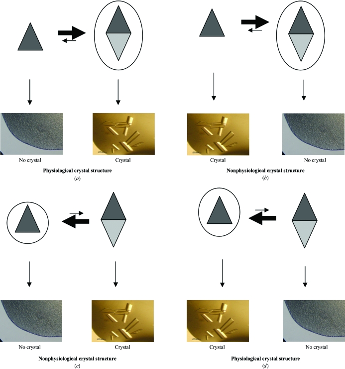
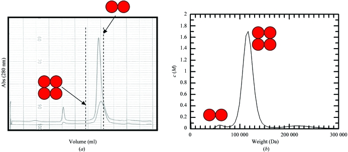
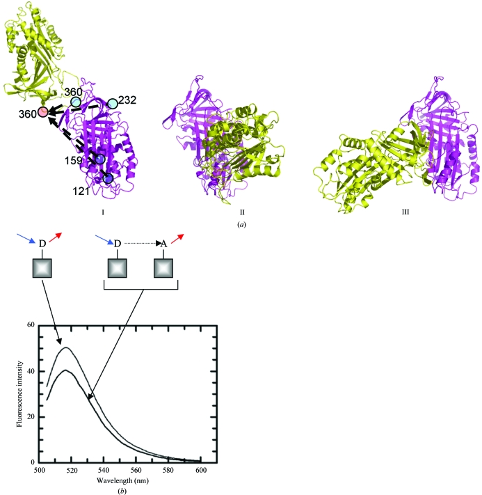
Protein in crystal form is at an extremely high concentration and yet retains the complex secondary structure that defines an active protein. The protein crystal itself is made up of a repeating lattice of protein-protein and protein-solvent interactions. The problem that confronts any crystallographer is to identify those interactions that represent physiological interactions and those that do not. This review explores the tools that are available to provide such information using the original crystal liquor as a sample. The review is aimed at postgraduate and postdoctoral researchers who may well be coming up against this problem for the first time. Techniques are discussed that will provide information on the stoichiometry of complexes as well as low-resolution information on complex structure. Together, these data will help to identify the physiological complex.
Figures
Similar articles
-
Reversible lattice repacking illustrates the temperature dependence of macromolecular interactions.J Mol Biol. 2001 Aug 24;311(4):851-62. doi: 10.1006/jmbi.2001.4891. J Mol Biol. 2001. PMID: 11518535
-
Softer and soft X-rays in macromolecular crystallography.J Synchrotron Radiat. 2005 Jul;12(Pt 4):410-9. doi: 10.1107/S0909049504025762. Epub 2005 Jun 15. J Synchrotron Radiat. 2005. PMID: 15968116 Review.
-
Advances in experimental phasing.Acta Crystallogr D Struct Biol. 2016 Mar;72(Pt 3):291-2. doi: 10.1107/S2059798316003375. Epub 2016 Mar 1. Acta Crystallogr D Struct Biol. 2016. PMID: 26960115 Free PMC article.
-
Towards an understanding of radiation damage in cryocooled macromolecular crystals.J Synchrotron Radiat. 2005 May;12(Pt 3):257-60. doi: 10.1107/S0909049505007132. Epub 2005 Apr 14. J Synchrotron Radiat. 2005. PMID: 15840908
-
Cryocooling and radiation damage in macromolecular crystallography.Acta Crystallogr D Biol Crystallogr. 2006 Jan;62(Pt 1):32-47. doi: 10.1107/S0907444905034207. Epub 2005 Dec 14. Acta Crystallogr D Biol Crystallogr. 2006. PMID: 16369092 Review.
Cited by
-
Evolutionary, physicochemical, and functional mechanisms of protein homooligomerization.Prog Mol Biol Transl Sci. 2013;117:3-24. doi: 10.1016/B978-0-12-386931-9.00001-5. Prog Mol Biol Transl Sci. 2013. PMID: 23663963 Free PMC article. Review.
-
The mitochondrial ADP/ATP carrier exists and functions as a monomer.Biochem Soc Trans. 2020 Aug 28;48(4):1419-1432. doi: 10.1042/BST20190933. Biochem Soc Trans. 2020. PMID: 32725219 Free PMC article. Review.
-
Characterization of the Molecular Weight of Hyaluronan in Eye Products Using a Novel Method of Size Exclusion High-Pressure Liquid Chromatography.Transl Vis Sci Technol. 2023 Apr 3;12(4):13. doi: 10.1167/tvst.12.4.13. Transl Vis Sci Technol. 2023. PMID: 37052911 Free PMC article.
-
Diffraction techniques in structural biology.Curr Protoc Nucleic Acid Chem. 2010 Jun;Chapter 7:Unit 7.13. doi: 10.1002/0471142700.nc0713s41. Curr Protoc Nucleic Acid Chem. 2010. PMID: 20517991 Free PMC article.
-
Diffraction Techniques in Structural Biology.Curr Protoc Nucleic Acid Chem. 2016 Jun 1;65:7.13.1-7.13.41. doi: 10.1002/cpnc.4. Curr Protoc Nucleic Acid Chem. 2016. PMID: 27248784 Free PMC article. Review.
References
-
- Bahadur, R. P., Chakrabarti, P., Rodier, F. & Janin, J. (2004). J. Mol. Biol.336, 943–955. - PubMed
-
- Brown, W. (1993). Dynamic Light Scattering: The Method and Some Applications. Oxford: Clarendon Press.
-
- Cooper, E. A. & Knutson, K. (1995). Pharm. Biotechnol.7, 101–143. - PubMed
-
- Dafforn, T. R., Mahadeva, R., Elliott, P. R., Sivasothy, P. & Lomas, D. A. (1999). J. Biol. Chem.274, 9548–9555. - PubMed
-
- Gettins, P. G. & Olson, S. T. (2004). Methods, 32, 110–119. - PubMed
Publication types
MeSH terms
Substances
LinkOut - more resources
Full Text Sources
Other Literature Sources
