

doi: 10.1107/S0907444906022657.
Epub 2006 Dec 13.
Crystallographic refinement of ligand complexes
Affiliations
- PMID: 17164531
- PMCID: PMC2483469
- DOI: 10.1107/S0907444906022657
Item in Clipboard
Crystallographic refinement of ligand complexes
Acta Crystallogr D Biol Crystallogr.
2007 Jan.
Abstract
Model building and refinement of complexes between biomacromolecules and small molecules requires sensible starting coordinates as well as the specification of restraint sets for all but the most common non-macromolecular entities. Here, it is described why this is necessary, how it can be accomplished and what pitfalls need to be avoided in order to produce chemically plausible models of the low-molecular-weight entities. A number of programs, servers, databases and other resources that can be of assistance in the process are also discussed.
Figures
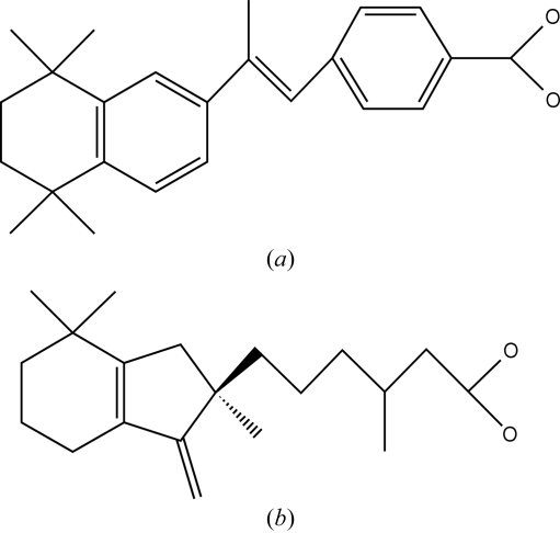
Chemical structure diagrams of (a) TTNPB and (b) ‘compound 19’.
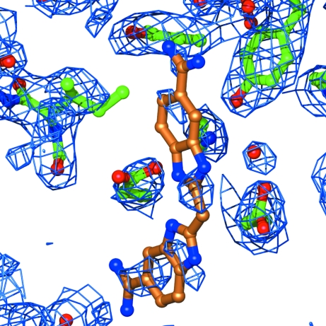
Electron density for the inhibitor BABIM (shown with gold C atoms) in its complex with botulinum neurotoxin type B protease (Hanson et al., 2000, 2002 ▶). The map is a 2mF
o − DF
c synthesis, calculated with all deposited data (2.5 Å), and taken from EDS (Kleywegt et al., 2004 ▶). Figs. 2 and 3 were created with O (Jones et al., 1991 ▶) and MolRay (Harris & Jones, 2001 ▶).
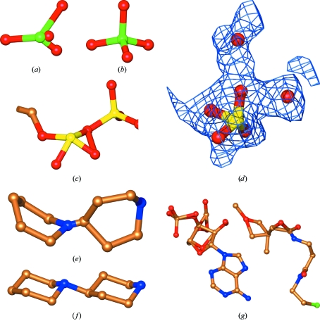
Examples of errors in heterocompounds encountered in contemporary wwPDB entries. (a) A sulfate ion as found in a 1.65 Å structure from 1999. One of the O atoms lies in an obviously impossible location. (b) Geometry of an ‘ideal’ sulfate from MSDChem. (c) Detail of an FAD molecule found in a 2.3 Å structure from 2005. One of the two phosphates has been subjected to incorrect restraints (in both copies in the asymmetric unit), forcing it into a tetragonal pyramidal structure. The other phosphate has its neighbouring O atoms in the proper tetrahedral arrangement. (d) A different, but equally wrong, phosphate. This 2.0 Å structural genomics structure from 2002 contains a phosphate forced into a trigonal pyramidal arrangement, with all four P—O bonds shorter than 1.5 Å (suggesting, incorrectly, that all four are double bonds). In the vicinity of this phosphate there is a large unoccupied density feature that looks as if it could also accommodate a phosphate ion (not shown). A nearby residue has density features that show that its peptide bond needs to be flipped (not shown). These uninterpreted yet obvious density features suggest that the maps have not been inspected with a great degree of enthusiasm. (e) The N atom in this ligand (found in a 2.5 Å structure from 2001) appears to have been forced to be planar. In addition, the bond from the N to the C atom in the other ring is implausibly short (0.8 Å). (f) The ‘ideal’ structure of the ligand in (e), taken from MSDChem. The r.m.s. deviation from ideal values of the bond lengths in the experimental structure is 0.2 Å and the r.m.s. deviation of the angles is 8°. (g) This poor impersonation of a coenzyme A molecule is found in a 2.25 Å structure from 2003. It contains non-bonded distances as short as 0.54 Å, bonded distances as long as 6.7 Å and bond angles as small as 18°.
References
-
- Aalten, D. M. F. van, Bywater, R., Findlay, J. B. C., Hendlich, M., Hooft, R. W. W. & Vriend, G. (1996). J. Comput. Aided Mol. Des.10, 255–262. - PubMed
-
- Berman, H., Henrick, K. & Nakamura, H. (2003). Nature Struct. Biol.10, 980. - PubMed
-
- Bohne, A., Lang, E. & von der Lieth, C. W. (1999). Bioinformatics, 15, 767–768. - PubMed
-
- Boström, J. (2001). J. Comput. Aided Mol. Des.15, 1137–1152. - PubMed
-
- Brünger, A. T. (1992). X-PLOR. Version 3.1. A System for X-ray Crystallography and NMR. Yale University, Connecticut, USA.
Publication types
MeSH terms
Substances
LinkOut - more resources
Full Text Sources
Other Literature Sources
