. 2006 Dec 13:6:26.
doi: 10.1186/1472-6807-6-26.

Structure of the yeast histone H3-ASF1 interaction: implications for chaperone mechanism, species-specific interactions, and epigenetics

Affiliations

Structure of the yeast histone H3-ASF1 interaction: implications for chaperone mechanism, species-specific interactions, and epigenetics

Andrew J Antczak et al. BMC Struct Biol. .

Abstract

Background: The histone H3/H4 chaperone Asf1 (anti-silencing function 1) is required for the establishment and maintenance of proper chromatin structure, as well as for genome stability in eukaryotes. Asf1 participates in both DNA replication-coupled (RC) and replication-independent (RI) histone deposition reactions in vitro and interacts with complexes responsible for both pathways in vivo. Asf1 is known to directly bind histone H3, however, high-resolution structural information about the geometry of this interaction was previously unknown.

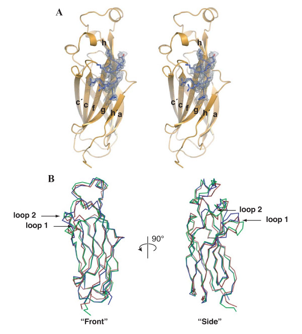
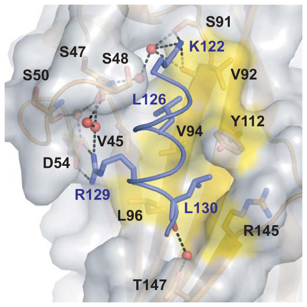
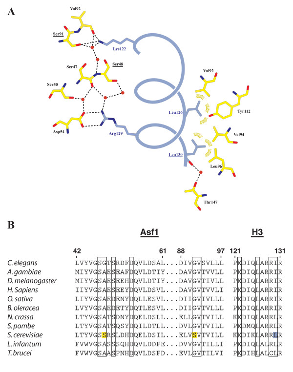
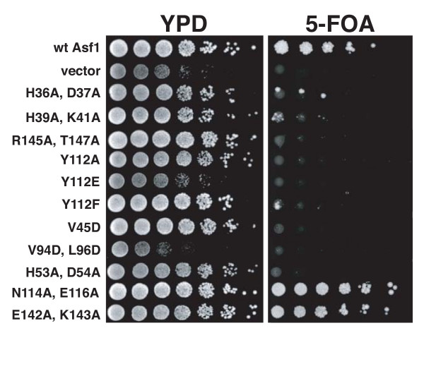
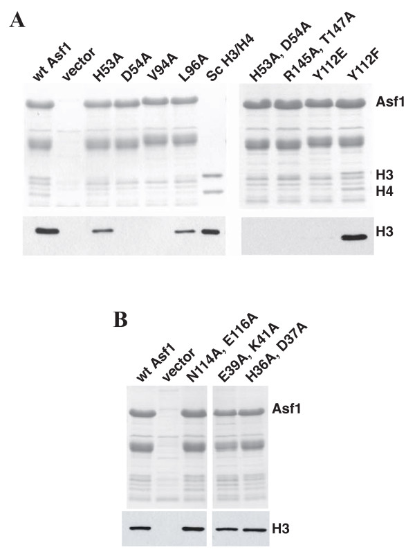
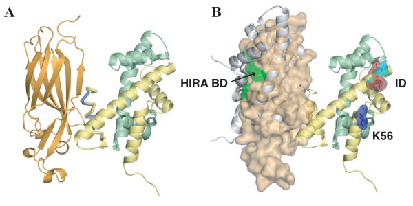
Results: Here we report the structure of a histone/histone chaperone interaction. We have solved the 2.2 A crystal structure of the conserved N-terminal immunoglobulin fold domain of yeast Asf1 (residues 2-155) bound to the C-terminal helix of yeast histone H3 (residues 121-134). The structure defines a histone-binding patch on Asf1 consisting of both conserved and yeast-specific residues; mutation of these residues abrogates H3/H4 binding affinity. The geometry of the interaction indicates that Asf1 binds to histones H3/H4 in a manner that likely blocks sterically the H3/H3 interface of the nucleosomal four-helix bundle.

Conclusion: These data clarify how Asf1 regulates histone stoichiometry to modulate epigenetic inheritance. The structure further suggests a physical model in which Asf1 contributes to interpretation of a "histone H3 barcode" for sorting H3 isoforms into different deposition pathways.


Figures

Figure 1
Figure 1
Global structure of Asf1N bound to H3α3. (A) Stereo view of Asf1N (orange) bound to the H3α3 helix (blue). β-strands are labeled as per the nomenclature for switched-type Ig-like folds. A section of an electron density omit-map generated in the absence of the histone helix is shown contoured at 2.5σ. (B) Front and side views superposing known Asf1 structures. α-carbon atoms of Asf1 from the current structure (green), the non-histone bound yeast Asf1 (PDB ID 1ROC, blue), and the human Asf1a (PDB ID 1TEY, red) were used for the alignment and shown as a-carbon representations. Arrows indicate the loops connecting b-strands c and c' (loop 1), and connecting b-strands e and f (loop 2), which both rotate forward upon binding H3α3.
Figure 2
Figure 2
Close-up view of the histone-binding patch on Asf1. Residues shown to be important for the interaction are labeled and represented as sticks, with labels in black for Asf1 and blue for histone H3 residues. Waters are represented as red spheres, hydrogen bonds as black dashed lines, and the hydrophobic surface participating in van der Waals interactions is shaded yellow.
Figure 3
Figure 3
Structural details of the Asf1N-H3a3 interface. (A) Schematic of the interactions between Asf1 and H3α3. Asf1 residues are displayed in yellow, H3α3 in blue. Yeast-specific residues on Asf1 and H3α3 are underlined. Ion pair and hydrogen bonding interactions measuring 3.3 Å or less are represented as dashed lines. Van der Waals interactions are shown as gold arcs. (B) Alignment of Asf1 and H3 residues involved in the interaction across several eukaryotes. Residues participating in the interaction are boxed, with yeast specific Asf1 residues highlighted in yellow and H3-Leu130 highlighted in blue. ClustalW was used to generate the Asf1 alignment.
Figure 4
Figure 4
Genetic analysis of Asf1 residues important for histone binding. Asf1 histone binding mutants cause silencing defects in vivo. cac1Δasf1Δ cells were transformed with plasmids expressing the indicated Asf1 proteins. A four-fold dilution series was spotted onto rich media (YPD) to indicate cell number and onto media containing 5-FOA to measure silencing of a telomeric URA3 reporter gene.
Figure 5
Figure 5
Biochemical analysis of Asf1 residues important for histone binding. (A) Asf1 proteins containing mutations in the H3 binding surface fail to pull-down histones H3/H4. Extracts were made from E. coli cells expressing yeast histones H3 and H4, as well as either wild type or mutant His6-tagged Asf1. Talon metal affinity resin was used to precipitate Asf1 and associated proteins. Samples were separated on a 17% SDS-PAGE gel, visualized by Coomassie staining to indicate recovery of Asf1 (upper panel), or analyzed by Western blotting using an anti-H3 antibody (AbCam) (lower panel). Sc H3/H4 indicates recombinant yeast histones H3/H4 (0.5 μg upper gel; 10 ng lower gel). (B) Mutation of Asf1 residues outside of the histone-binding patch does not disrupt its interaction with histones H3/H4. The experiment was performed as above.
Figure 6
Figure 6
Model of H3/H4 dimer binding by Asf1N. (A) Asf1 binds to a dimer of histones H3/H4. The structure of Asf1N-H3α3 was modeled onto the structure of the yeast nucleosome (PDB ID 1ID3) by aligning the α-carbons of H3α3 with the respective α-carbons of the nucleosome structure. Asf1 is shown in orange and is represented in cartoon form. Nucleosomal histones H3 (yellow) and H4 (green) are shown in cartoon form with the H3a3-helix represented as a Cα trace for the nucleosomal (yellow) and complex crystal structure (blue). (B) Asf1 occludes binding of a second H3/H4 dimer while leaving critical decoding residues surface exposed. Asf1 is shown with a transparent surface representation. An H3/H4 dimer that would be occluded by Asf1 binding is shown as a grey cartoon. Yeast (light blue) and metazoan (red) H3 residues that differ among the H3.1/H3.2 and H3.3 isoforms are shown as sticks with a transparent surface representation, and labeled "ID" for "Isoform Determinants". The H3-K56 residue (dark blue) that is acetylated on newly synthesized histones and the region responsible for HIRA binding (green, labeled "HIRA BD"), are also shown.

References

    1. Woodcock CL. Chromatin architecture. Curr Opin Struct Biol. 2006;16:213–220. doi: 10.1016/j.sbi.2006.02.005. - DOI - PubMed
    1. Shibahara K, Stillman B. Replication-dependent marking of DNA by PCNA facilitates CAF-1-coupled inheritance of chromatin. Cell. 1999;96:575–585. doi: 10.1016/S0092-8674(00)80661-3. - DOI - PubMed
    1. Ray-Gallet D, Quivy JP, Scamps C, Martini EM, Lipinski M, Almouzni G. HIRA is critical for a nucleosome assembly pathway independent of DNA synthesis. Mol Cell. 2002;9:1091–1100. doi: 10.1016/S1097-2765(02)00526-9. - DOI - PubMed
    1. Green EM, Antczak AJ, Bailey AO, Franco AA, Wu KJ, Yates JR, 3rd, Kaufman PD. Replication-independent histone deposition by the HIR complex and Asf1. Curr Biol. 2005;15:2044–2049. doi: 10.1016/j.cub.2005.10.053. - DOI - PMC - PubMed
    1. Prochasson P, Florens L, Swanson SK, Washburn MP, Workman JL. The HIR corepressor complex binds to nucleosomes generating a distinct protein/DNA complex resistant to remodeling by SWI/SNF. Genes Dev. 2005;19:2534–2539. doi: 10.1101/gad.1341105. - DOI - PMC - PubMed

Publication types

MeSH terms

Substances

Associated data

LinkOut - more resources