. 2007 Apr 25;361(1):212-28.
doi: 10.1016/j.virol.2006.11.004. Epub 2006 Dec 12.

HIV-1 clones resistant to a small molecule CCR5 inhibitor use the inhibitor-bound form of CCR5 for entry

Affiliations

HIV-1 clones resistant to a small molecule CCR5 inhibitor use the inhibitor-bound form of CCR5 for entry

Pavel Pugach et al. Virology. .

Abstract

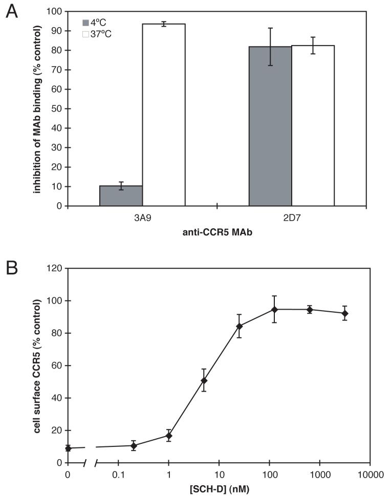
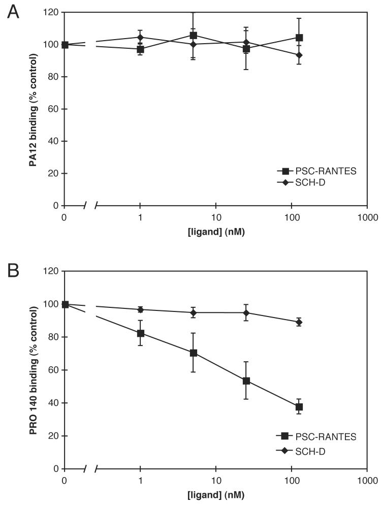
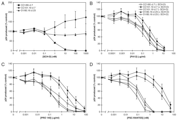
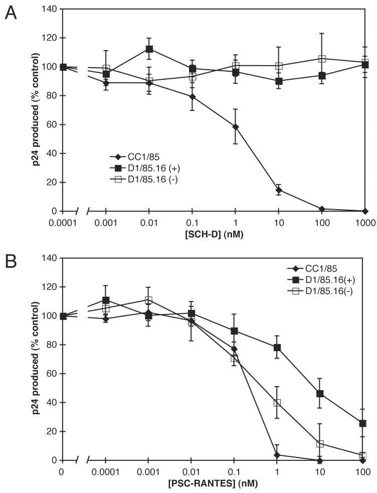
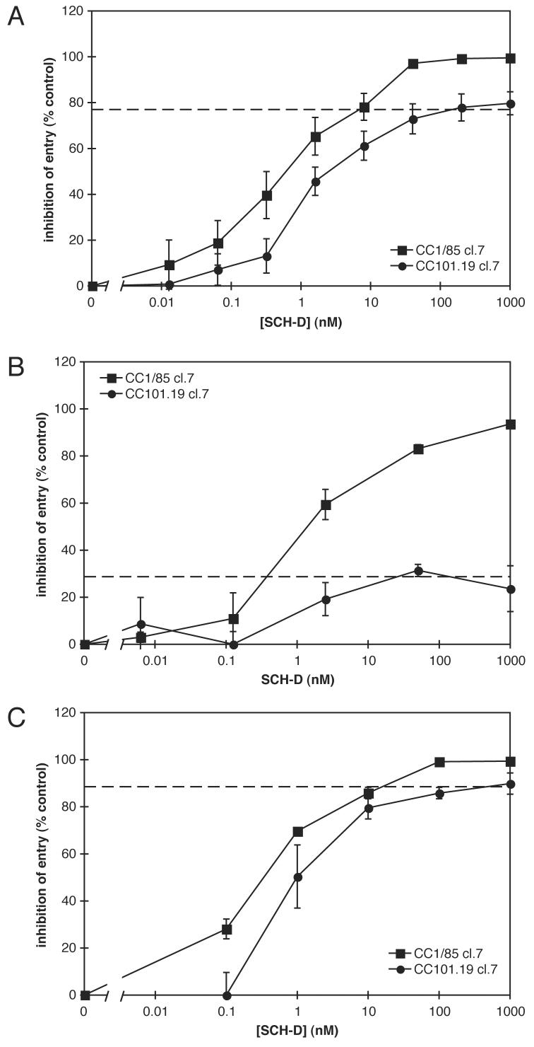
Human immunodeficiency virus type 1 (HIV-1) infection can be inhibited by small molecules that target the CCR5 coreceptor. Here, we describe some properties of clonal viruses resistant to one such inhibitor, SCH-D, using both chimeric, infectious molecular clones and Env-pseudotypes. Studies using combinations of CCR5 ligands, including small molecule inhibitors, monoclonal antibodies (MAbs) and chemokine derivatives such as PSC-RANTES, show that the fully SCH-D-resistant viruses enter target cells by using the SCH-D-bound form of CCR5. However, the way resistance to SCH-D and other small molecule CCR5 inhibitors is manifested depends on the target cell and the nature of the assay (single- vs. multi-cycle). In multi-cycle assays using primary lymphocytes, SCH-D does not inhibit resistant molecular clones, and it can even enhance their infectivity modestly. In contrast, the same viruses (as Env-pseudotypes) are significantly inhibited by SCH-D in single-cycle entry assays using U87-CD4/CCR5 cells, resistance being manifested by incomplete inhibition at high SCH-D concentrations. When a single-cycle, Env-pseudotype entry assay was performed using either U87-CD4/CCR5 cells or PBMC under comparable conditions, entry was inhibited by up to 88% in the former cells but by only 28% in the PBMC. Hence, there are both cell- and assay-dependent influences on how resistance is manifested. We also take this opportunity to correct our previous report that SCH-D-resistant isolates are also substantially cross-resistant to PSC-RANTES [Marozsan, A.J., Kuhmann, S.E., Morgan, T., Herrera, C., Rivera-Troche, E., Xu, S., Baroudy, B.M., Strizki, J., Moore, J.P., 2005. Generation and properties of a human immunodeficiency virus type 1 isolate resistant to the small molecule CCR5 inhibitor, SCH-417690 (SCH-D). Virology 338 (1), 182-199]. A substantial element of this resistance was attributable to the unappreciated carry-over of SCH-D from the selection cultures into analytical assays.


Figures

Figure 1
Figure 1
SCH-D is an antagonist of the PSC-RANTES induced internalization. A. HeLa-CD4/CCR5 cells were incubated for 1 h at 4°C (shaded bars) or 37°C (open bars) with or without 300 nM PSCRANTES. The cells were then stained for 1 h at 4°C with PE-labeled 3A9 or 2D7. The data shown represent the inhibition of MAb binding after incubation with 300 nM PSC-RANTES relative to cells stained after incubation in the absence of PSC-RANTES. The mean values ± the standard error of the mean (SEM), derived from three independent experiments, are shown. B. The effects of varying SCH-D concentrations on PSC-RANTES-induced CCR5 internalization were determined. HeLa-CD4/CCR5 cells were incubated for 1 h at 37°C with SCH-D at the indicated concentration before the addition of 300 nM PSC-RANTES for 1 h at 37°C. The cells were then stained at 4°C for 1 h with PE-labeled 3A9. The CCR5 expression level was calculated as a percent of the specific 3A9 staining in the same experiment under the same conditions but without PSC-RANTES addition. The data points are the means of between 3 and 7 independent experiments at each SCH-D concentration, and are shown ± SEM.
Figure 2
Figure 2
How SCH-D and PSC-RANTES interact with PRO 140 and PA12. HeLa-CD4/CCR5 cells were incubated with the indicated concentrations of SCH-D (diamonds) or PSC-RANTES (squares) for 1 h at 4°C, and then with 25 μg/ml PA12 (A) or 30 μg/ml PRO 140 (B) at 4°C for 1 h. Bound MAbs were detected with a PE-labeled anti-mouse IgG1 MAb (A) or a FITC-labeled anti-human IgG4 MAb (B). In both panels, the specific MFI values are expressed as a percentage of those determined under the same conditions, but in the absence of SCH-D or PSC-RANTES, and are the means of three independent experiments ± SEM.
Figure 3
Figure 3
Effects of CCR5 inhibitors alone or in combination with SCH-D on replication of clonal viruses in PBMC. A. PBMC were incubated with the indicated concentration of SCH-D for 1 h at 37°C before the addition of the CC1/85 cl.7 (squares), CC101.19 cl.7 (circles) or D1/85.16 cl.23 (triangles) clonal viruses. After 7 days of culture the production of p24 antigen under each condition was assessed by ELISA. The results show p24 production as a percentage of that derived from cells that were not treated with SCH-D. The values shown are the means ± SEM from 6 independent experiments. B-D. PBMC were incubated with (closed symbols) or without (open symbols) 1 μM SCH-D for 1 h prior to the addition of the indicated concentrations of PA12 (B), PRO 140 (C) or PSC-RANTES (D) for 1 h at 37°C. The CC1/85 cl.7 (squares), CC101.19 cl.7 (circles) or D1/85.16 cl.23 (triangles) clonal viruses were then added. After 7 days of culture p24 production under each condition was assessed by ELISA. The results show p24 production as a percentage of that produced by cells infected in the absence of PA12, PRO 140 or PSC-RANTES, but in the presence (closed symbols) or absence (open symbols) of 1 μM SCH-D. The data for CC1/85 cl.7 in the presence of 1 μM SCH-D could, therefore, not be plotted (no p24 was produced even in the absence of another inhibitor). The addition of PA12, PRO 140 or PSC-RANTES did not reverse the lack of p24 production by CC1/85 cl.7 in the presence of 1 μM SCHD. The values shown are the means ± SEM from 6 or 7 independent experiments.
Figure 4
Figure 4
The effect of residual SCH-D on the PSC-RANTES resistance of SCH-D-resistant isolates. PBMC were incubated with the indicated concentration of SCH-D (A) or PSC-RANTES (B) for 1 h at 37°C before the addition of virus isolates CC1/85 (diamonds) or D1/85.16 (squares). Open symbols represent D1/85.16 isolate from which residual SCH-D had been thoroughly removed, whereas filled symbols represent D1/85.16 isolate that was harvested in the presence of 25 μM SCH-D. After 7 days of culture, the amount of p24 antigen produced under each condition was assessed by ELISA. The results show p24 production as a percentage of that produced by cells infected in the absence of any CCR5 inhibitor. The values shown are the means ± SEM from 4 or 5 independent experiments.
Figure 5
Figure 5
Single-round infection assays of SCH-D sensitivity of the CC1.85 cl.7 and CC101.19 cl.7 Envs. A. U87-CD4/CCR5 cells were incubated with the indicated concentration of SCH-D for 1 h at 37°C before the addition of luciferase-transducing viruses pseudotyped with the CC1/85 cl.7 (squares) or CC101.19 cl.7 (circles) Env proteins. After three days of incubation, the cells were assayed for luciferase activity. The results are shown as percent inhibition where 0% inhibition is defined as the luciferase activity in cells infected in the absence of SCH-D and 100% inhibition is defined as the luciferase activity measured in cells that were not infected with the pseudoviruses. The values shown are the means ± SEM from 5 independent experiments. The dashed line indicates the apparent plateau in the CC101.19 cl.7 results. B. and C. PBMC (B) or U87-CD4/CCR5 cells (C) were incubated with the indicated concentration of SCH-D for 1 h at 37°C before the addition of hrGFPII-transducing viruses pseudotyped with the CC1/85 cl.7 (squares) or CC101.19 cl.7 (circles) Env proteins. After four days of incubation, the cells were assayed by cytometry for hrGFPII expression. The results are shown as percent inhibition where 0% inhibition is defined as the fraction of hrGFPII+ cells in the absence of SCH-D and 100% inhibition is defined as no hrGFPII+ cells. The values shown are the means ± SEM from 3 independent experiments. The dashed lines indicate the apparent plateau in the CC101.19 cl.7 results.
Figure 6
Figure 6
Changes in CC-chemokine production from, and CCR5 expression on, PBMC treated with CCR5 antagonists. A. Stimulated PBMC from seven individuals were incubated for seven days with or without PRO 140 (50 μg/ml) or SCH-D (2 μM) as indicated. The culture supernatants were assayed for MIP-1α (white bars), MIP-1β (grey bars) or RANTES (black bars) content. The data shown are the mean values ± SEM for the fold-increases, compared to no treatment, from the seven donors. B. In the same experiment as in (A), stimulated PBMC from the same seven individuals were incubated for seven days with or without SCH-D as indicated. CD4 expression was assessed with PerCP-labeled anti-CD4, CCR5 expression using the PE-labeled anti-CCR5 MAb 2D7; both the percentage of CD4+ T-cells that were also CCR5+ and the mean fluorescence intensity (MFI) for the CD4+CCR5+ T-cells were determined, as indicated. The data shown are the mean values ± SEM for the fold-increases, compared to no treatment, from the seven donors.
Figure 7
Figure 7
Schematic depiction of possible mechanisms of resistance to an allosteric small-molecule CCR5 inhibitor. A. CCR5 is depicted with two distinct HIV-1 interaction sites (peaks) and a separate SCH-D (or other allosteric CCR5 inhibitor) binding site (valley). The gp120 protein is depicted as having two interaction sites that are compatible with binding to CCR5, thereby mediating infection. For convenience in drawing the figure, we depict the gp120-CCR5 interaction via one of these sites as being weaker than the other, leaving room for a stronger interaction in (D). This need not necessarily be the case, as the strengthened interaction in (D) could involve both interaction sites. B. In the presence of a high SCH-D concentration, the conformation of one of the interaction regions on CCR5 is altered, prohibiting interaction with gp120 and preventing infection. C. Noncompetitive resistance is depicted as a change in the conformation of gp120 to accommodate the altered CCR5 conformation. In the case of the noncompetitive resistance described in this paper, infection through the SCH-D-free form of CCR5 is also possible for gp120 from the SCH-D-resistant viruses we have studied here. How this form of resistance would be manifested in an entry assay at varying efficiencies of entry through the SCH-DCCR5 complex (relative to free CCR5) is shown below the diagram. D. Competitive resistance is depicted as a change in the conformation of gp120 to increase the affinity of gp120 for CCR5 (here shown by a better fit between the two). In this scenario, gp120 better competes with SCH-D for binding to CCR5. How this form of resistance would be manifested in an entry assay with various degrees of improvement in the gp120-CCR5 interaction (relative to the wild type gp120) is shown below the diagram.

References

    1. Billick E, Seibert C, Pugach P, Ketas T, Trkola A, Endres MJ, Murgolo NJ, Coates E, Reyes GR, Baroudy BM, Sakmar TP, Moore JP, Kuhmann SE. The differential sensitivity of human and rhesus macaque CCR5 to small-molecule inhibitors of human immunodeficiency virus type 1 entry is explained by a single amino acid difference and suggests a mechanism of action for these inhibitors. J. Virol. 2004;78(8):4134–4144. - PMC - PubMed
    1. Bjorndal A, Deng H, Jansson M, Fiore JR, Colognesi C, Karlsson A, Albert J, Scarlatti G, Littman DR, Fenyo EM. Coreceptor usage of primary human immunodeficiency virus type 1 isolates varies according to biological phenotype. J. Virol. 1997;71(10):7478–7487. - PMC - PubMed
    1. Coakley E, Petropoulos CJ, Whitcomb JM. Assessing chemokine co-receptor usage in HIV. Curr. Opin. Infect. Dis. 2005;18(1):9–15. - PubMed
    1. Cormier EG, Dragic T. The crown and stem of the V3 loop play distinct roles in human immunodeficiency virus type 1 envelope glycoprotein interactions with the CCR5 coreceptor. J. Virol. 2002;76(17):8953–8957. - PMC - PubMed
    1. Cormier EG, Tran DN, Yukhayeva L, Olson WC, Dragic T. Mapping the determinants of the CCR5 amino-terminal sulfopeptide interaction with soluble human immunodeficiency virus type 1 gp120-CD4 complexes. J. Virol. 2001;75(12):5541–5549. - PMC - PubMed

Publication types