. 2007 Mar;81(5):2158-64.
doi: 10.1128/JVI.02070-06. Epub 2006 Dec 13.

Basolateral entry and release of Crimean-Congo hemorrhagic fever virus in polarized MDCK-1 cells

Affiliations

Basolateral entry and release of Crimean-Congo hemorrhagic fever virus in polarized MDCK-1 cells

Anne-Marie Connolly-Andersen et al. J Virol. 2007 Mar.

Abstract

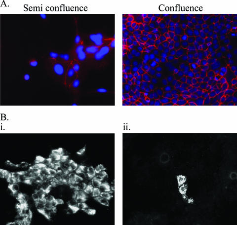
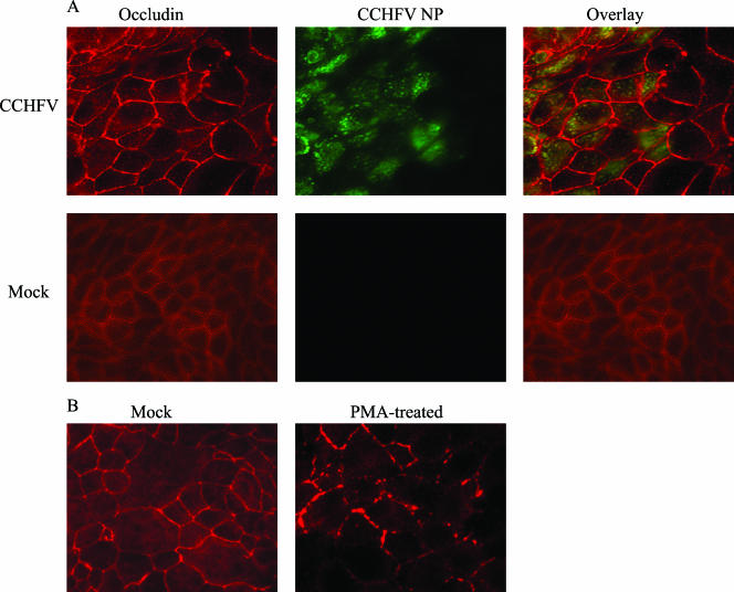
Crimean-Congo hemorrhagic fever virus (CCHFV) is an etiological agent of a disease with mortality rates in patients averaging 30%. The disease is characterized by fever, myalgia, and hemorrhage. Mechanisms underlying the hemorrhage have to our knowledge not been elucidated for CCHFV. Possibly, a direct or indirect viral effect on tight junctions (TJ) could cause the hemorrhage observed in patients, as TJ play a crucial role in vascular homeostasis and can cause leakage upon deregulation. Moreover, there is no knowledge regarding the site of entry and release of CCHFV in polarized epithelial cells. Such cells represent a barrier to virus dissemination within the host, and as a site of viral entry and release, they could play a key role in further spread. For the first time, we have shown preferential basolateral entry of CCHFV in Madin-Darby canine kidney 1 (MDCK-1) epithelial cells. Furthermore, we demonstrated basolateral release of CCHFV in polarized epithelial cells. Interestingly, by measuring transepithelial electrical resistance, we found no effect of CCHFV replication on the function of TJ in this study. Neither did we observe any difference in the localization of the TJ proteins ZO-1 and occludin in CCHFV-infected cells compared to mock-infected cells.


Figures

FIG. 1.
FIG. 1.
(A) Semiconfluent cells are not polarized as indicated by TJ staining. Semiconfluent and confluent cell layers were stained for occludin (red) in order to indicate the level of cell polarization upon infection with CCHFV. Cell nucleus is indicated by DAPI staining (blue). (B) Subconfluent cell layers are more susceptible to infection with CCHFV than confluent cell layers. MDCK-1 cell layers were grown to either semiconfluence (i) or confluence (ii) and inoculated with CCHFV at an MOI of 0.02. Cell layers were fixed and stained for CCHFV NP 48 hpi and examined by immunofluorescence microscopy.
FIG. 2.
FIG. 2.
CCHFV has basolateral entry. (A) MDCK-1 cells were grown to confluence on Costar membranes, and a viral suspension was added to either the basolateral or the apical compartment at an MOI of 1. At 72 hpi, the cells were harvested and analyzed for intracellular viral nucleocapsids by Western blotting using calnexin as a loading reference. (B) MDCK-1 cells were infected apically at semiconfluence and confluence as indicated by TER and thereafter infected with CCHFV at an MOI of 1. At 48 hpi, cells were harvested and analyzed for infectivity by detecting viral nucleocapsids by Western blotting. (C) Confluent cell layers were treated or mock treated with 15 mM EGTA for 1 h and thereafter apically infected with CCHFV at an MOI of 1. At 48 hpi, the cells were harvested and CCHFV NP was detected by Western blotting.
FIG. 3.
FIG. 3.
CCHFV progeny virion release is primarily basolateral. Confluent monolayers on semipermeable membranes were inoculated basolaterally with CCHFV at an MOI of 1. Infected supernatants were retrieved daily and exchanged for fresh medium. The titers of infectious virions in the basolateral and apical supernatants (sup) were determined by fluorescence focus unit assay as described in Materials and Methods. Values from TER are given to indicate monolayer integrity for both mock-infected cells and infected cells.
FIG. 4.
FIG. 4.
The integrity of TJ is not disturbed by CCHFV. (A) Confluent cell layers grown on 3.0-μm semipermeable membranes were inoculated with an MOI of 1 of CCHFV either apically or basolaterally. TER was measured daily, and results are mean values of triplicates. (B) PMA at 100 and 1,000 nM was added to confluent cell layers on 3.0-μm-pore membranes, and TER was measured every 30 min. The results are mean values of triplicates.
FIG. 5.
FIG. 5.
Occludin localizes to cell-cell boundaries in infected MDCK-1 cells. (A) Subconfluent cells grown in chamber slides were either infected with CCHFV (top row) or mock infected (bottom row) and fixed 48 hpi. CCHFV NP is shown in green, and occludin is shown in red. (B) Confluent cells were mock treated or treated with PMA (100 nM) for 1 h. After fixation, cells were stained for occludin as described in Materials and Methods.
FIG. 6.
FIG. 6.
ZO-1 localizes to cell-cell boundaries in infected MDCK-1 cells. (A) Subconfluent cell layers of MDCK-1 in chamber slides were either infected with CCHFV (top row) or mock infected (bottom row) and fixed 48 hpi. CCHFV NP is shown in red, and ZO-1 is shown in green. (B) Confluent cells were mock treated or treated with PMA (100 nM) for 1 h. After fixation, cells were stained for ZO-1 as described in Materials and Methods.

References

    1. Andersson, I., M. Simon, A. Lundkvist, M. Nilsson, A. Holmstrom, F. Elgh, and A. Mirazimi. 2004. Role of actin filaments in targeting of Crimean Congo hemorrhagic fever virus nucleocapsid protein to perinuclear regions of mammalian cells. J. Med. Virol. 72:83-93. - PubMed
    1. Bazzoni, G., and E. Dejana. 2004. Endothelial cell-to-cell junctions: molecular organization and role in vascular homeostasis. Physiol. Rev. 84:869-901. - PubMed
    1. Bertolotti-Ciarlet, A., J. Smith, K. Strecker, J. Paragas, L. A. Altamura, J. M. McFalls, N. Frias-Staheli, A. Garcia-Sastre, C. S. Schmaljohn, and R. W. Doms. 2005. Cellular localization and antigenic characterization of Crimean-Congo hemorrhagic fever virus glycoproteins. J. Virol. 79:6152-6161. - PMC - PubMed
    1. Bosch, I., K. Xhaja, L. Estevez, G. Raines, H. Melichar, R. V. Warke, M. V. Fournier, F. A. Ennis, and A. L. Rothman. 2002. Increased production of interleukin-8 in primary human monocytes and in human epithelial and endothelial cell lines after dengue virus challenge. J. Virol. 76:5588-5597. - PMC - PubMed
    1. Bray, M., and T. W. Geisbert. 2005. Ebola virus: the role of macrophages and dendritic cells in the pathogenesis of Ebola hemorrhagic fever. Int. J. Biochem. Cell Biol. 37:1560-1566. - PubMed

Publication types

LinkOut - more resources