. 2007 Mar;81(5):2231-9.
doi: 10.1128/JVI.01979-06. Epub 2006 Dec 13.

Degradation of tyrosine phosphatase PTPN3 (PTPH1) by association with oncogenic human papillomavirus E6 proteins

Affiliations

Degradation of tyrosine phosphatase PTPN3 (PTPH1) by association with oncogenic human papillomavirus E6 proteins

Ming Jing et al. J Virol. 2007 Mar.

Abstract

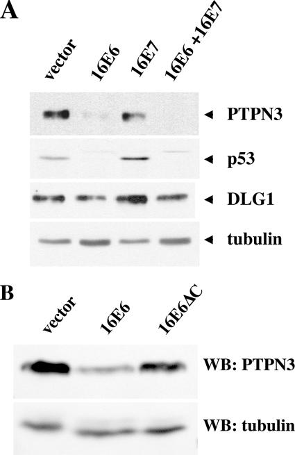
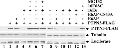
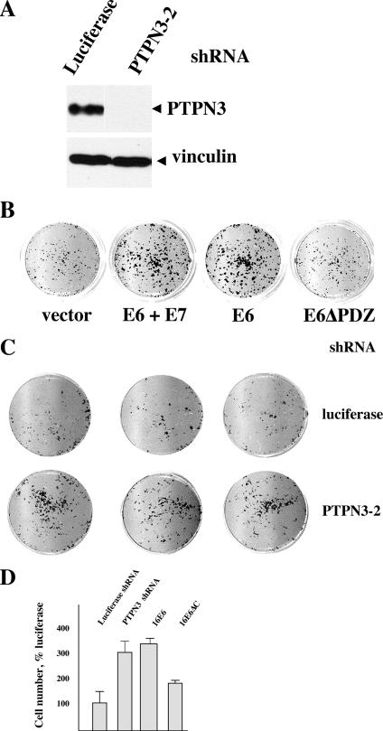
Oncoproteins from DNA tumor viruses associate with critical cellular proteins to regulate cell proliferation, survival, and differentiation. Human papillomavirus (HPV) E6 oncoproteins have been previously shown to associate with a cellular HECT domain ubiquitin ligase termed E6AP (UBE3A). Here we show that the E6-E6AP complex associates with and targets the degradation of the protein tyrosine phosphatase PTPN3 (PTPH1) in vitro and in living cells. PTPN3 is a membrane-associated tyrosine phosphatase with FERM, PDZ, and PTP domains previously implicated in regulating tyrosine phosphorylation of growth factor receptors and p97 VCP (valosin-containing protein, termed Cdc48 in Saccharomyces cerevisiae) and is mutated in a subset of colon cancers. Degradation of PTPN3 by E6 requires E6AP, the proteasome, and an interaction between the carboxy terminus of E6 and the PDZ domain of PTPN3. In transduced keratinocytes, E6 confers reduced growth factor requirements, a function that requires the PDZ ligand of E6 and that can in part be replicated by inhibiting the expression of PTPN3. This report demonstrates the potential of E6 to regulate phosphotyrosine metabolism through the targeted degradation of a tyrosine phosphatase.


Figures

FIG. 1.
FIG. 1.
16E6 recruits the cellular tyrosine phosphatase PTPN3 protein to E6AP. (A) 16E6 recruits a 128-kDa protein to the E6AP ubiquitin ligase. Ubiquitin ligase-defective E6AP (E6AP-C843A) tagged at the amino terminus with the EE and Flag epitopes was transiently expressed with or without untagged 16E6 in CV-1 cells. Cleared lysates were purified by tandem affinity purification with EE and Flag antibodies followed by peptide elution and analysis on 5 to 20% SDS-polyacrylamide silver-stained gels. Lanes 1 and 2, molecular weight markers; lane 4, tandem affinity-purified (TAP), vector-transfected cells; lane 6, TAP, EE-Flag-E6AP-C843A-transfected cells; lane 8, TAP EE-Flag-E6AP-C843A expressed together with untagged 16E6; lanes 3, 5, and 7, loaded with sample buffer only. Peptides of PTPN3 sequenced from p128 band numbered by GenBank NP_002820: FFIPDPNTLQQEQTR (amino acids [aa] 109 to 115), VESLHEQHSGLK (aa 188 to 199), TLDFYGVELHSGR (aa 213 to 225), EHIVAFNMLNYR (aa 281 to 292), SCVEHHTFFQAK (aa 300 to 311), LLPQEK (aa 313 to 318), NVLSQYWTMGSR (aa 319 to 330), ITPDEDGKFGFNLK (aa 513 to 526), MPLVVSR (aa 533 to 539), INPESPADTCIPK (aa 540 to 552), LNEGDQIVLINGR (aa 553 to 565), ELALVIR (aa 589 to 595), GLESGTVLIQFEQLYR (aa 640 to 655), KKPGLAITFAK (aa 656 to 666), LPQNLDKNR (aa 667 to 675), YKDVLPYDTTR (aa 657 to 686), MRDQRAMMVQTSSQYK (aa 877 to 892), and FVCEAILR (aa 893 to 900). The positions of molecular weight standards (in thousands) are shown to the left of the gel. (B) Domain structure of PTPN3. A diagram of PTPN3 illustrates the relative locations of FERM, PDZ, and phosphatase domains. Black squares illustrate the approximate location of sequenced peptides from mass spectrometry of p128. Below are diagrammed plasmids used in this study with the amino acid segments of PTPN3. WT, wild type; ins, inserted.
FIG. 2.
FIG. 2.
In vitro degradation of PTPN3 by high-risk mucosal E6 proteins. PTPN3, p53, and the indicated 16E6 types from cancer-associated HPV (16E6 and 18E6), non-cancer-associated mucosal HPV (11E6), non-cancer-associated cutaneous HPV (1E6), cancer-associated cutaneous HPV (8E6), and bovine papillomavirus type 1 (BE6) were in vitro translated in reticulocyte lysate containing 35S-labeled amino acids and incubated together for 60 min at room temperature to assay for in vitro degradation of 35S-labeled p53 and PTPN3. Samples were analyzed by SDS-PAGE, with the segments containing p53, PTPN3, and E6 proteins indicated (broadening of some E6 bands is due to comigration with globin from the reticulocyte lysate).
FIG. 3.
FIG. 3.
Role of PDZ interactions in the degradation of PTPN3 by 16E6. (A) An intact PDZ domain on PTPN3 is required for efficient binding to 16E6 in vitro. Input and GST-16E6 bound in vitro translation products of PTPN3 and the indicated mutants of PTPN3 (Fig. 1B) were resolved on SDS-polyacrylamide gels fluorographed and quantified by beta counting. GST-16E6 is shown from Coomassie blue staining. Bound counts resulting from four independent binding reactions are graphed below with standard deviation error bars. WT, wild type. (B) 16E6 degradation of PTPN3 requires the carboxy-terminal PDZ-binding peptide of 16E6. In vitro-translated p53, PTPN3, 16E6 or the indicated 16E6 mutants were mixed together as indicated and incubated for 60 min at 25°C. 16E6F2V refers to substitution of valine for phenylalanine at amino acid position 2, and 16E6ΔC contains a deletion of the carboxy-terminal 2 amino acids of 16E6. Aliquots removed at 0 and 60 min were analyzed by SDS-PAGE followed by autoradiography. (C) An intact PDZ domain in PTPN3 is required for in vitro degradation by 16E6. In vitro-translated p53, PTPN3, or the indicated PTPN3 mutants were incubated with mock-translated lysate,16E6, or 16E6ΔC for 60 min. (D) Comparison of in vitro degradation of PTPN3, p53, and DLG1. PTPN3, p53, and DLG1 were in vitro translated in reticulocyte lysate with 35S-labeled amino acids and incubated with in vitro-translated 16E6, 18E6, or 18E6ΔC for 0 or 60 min before analysis by SDS-PAGE and autoradiography. 16E6 bands are distorted by reticulocyte-derived globin in the gel.
FIG. 3.
FIG. 3.
Role of PDZ interactions in the degradation of PTPN3 by 16E6. (A) An intact PDZ domain on PTPN3 is required for efficient binding to 16E6 in vitro. Input and GST-16E6 bound in vitro translation products of PTPN3 and the indicated mutants of PTPN3 (Fig. 1B) were resolved on SDS-polyacrylamide gels fluorographed and quantified by beta counting. GST-16E6 is shown from Coomassie blue staining. Bound counts resulting from four independent binding reactions are graphed below with standard deviation error bars. WT, wild type. (B) 16E6 degradation of PTPN3 requires the carboxy-terminal PDZ-binding peptide of 16E6. In vitro-translated p53, PTPN3, 16E6 or the indicated 16E6 mutants were mixed together as indicated and incubated for 60 min at 25°C. 16E6F2V refers to substitution of valine for phenylalanine at amino acid position 2, and 16E6ΔC contains a deletion of the carboxy-terminal 2 amino acids of 16E6. Aliquots removed at 0 and 60 min were analyzed by SDS-PAGE followed by autoradiography. (C) An intact PDZ domain in PTPN3 is required for in vitro degradation by 16E6. In vitro-translated p53, PTPN3, or the indicated PTPN3 mutants were incubated with mock-translated lysate,16E6, or 16E6ΔC for 60 min. (D) Comparison of in vitro degradation of PTPN3, p53, and DLG1. PTPN3, p53, and DLG1 were in vitro translated in reticulocyte lysate with 35S-labeled amino acids and incubated with in vitro-translated 16E6, 18E6, or 18E6ΔC for 0 or 60 min before analysis by SDS-PAGE and autoradiography. 16E6 bands are distorted by reticulocyte-derived globin in the gel.
FIG. 4.
FIG. 4.
16E6 reduces the in vivo levels of PTPN3 in stably transduced cells. (A) 16E6 reduces PTPN3 expression in NIKS keratinocytes. NIKS human keratinocytes stably retrovirally transduced by the indicated oncogenes were removed from feeder cells, reattached to collagen type 1-coated plates overnight in 3T3 feeder cell-conditioned medium, and lysed in SDS lysis buffer the next day, and equalized amounts of protein were analyzed by sequential probing of a Western blot for the expression of PTPN3, p53, DLG1, and tubulin. (B) In vivo reduction of PTPN3 by 16E6 requires an intact PDZ motif on 16E6. HaCat keratinocytes stably transduced with the indicated retroviral plasmids were lysed in SDS sample buffer, and equal amounts of protein were analyzed with antibodies to PTPN3 (top panel) or tubulin (bottom panel). The blot exposures were quantitated by densitometry scanning to determine that the residual amounts of PTPN3 in lane 2 were 22% of lane 1 and 79% in lane 3. WB, Western blotting.
FIG. 5.
FIG. 5.
Role of E6AP in proteasome-mediated degradation of PTPN3 by 16E6. E6AP null mouse embryo fibroblasts were transfected with the indicated plasmids together with a constant amount of internal luciferase expression plasmid (starting in lane 2) and empty vector DNA to make the amount of transfected DNA constant. Cells were lysed in SDS sample buffer 20 h later and analyzed sequentially by immunoblotting with antibodies to FLAG, tubulin, and luciferase. PTPN3 was tagged at the carboxy-terminal end with the FLAG epitope. The results of one representative experiment of five experiments are shown.
FIG. 6.
FIG. 6.
Role of the PDZ ligand of E6 in reducing the growth factor requirements of keratinocytes. (A) shRNA to PTPN3 reduces PTPN3 expression. Epithelial cells transduced with the indicated retroviruses were lysed in SDS sample buffer and analyzed by immunoblotting for PTPN3 expression. Each lane shows the result of independently transduced cell cultures arising from pooled drug-resistant cell colonies. (B) Role of the E6 PDZ ligand in the acquisition of reduced growth factor requirements. A total of 5 × 10³ NIKS keratinocytes transduced by the indicated retroviruses were seeded into wells with mitomycin C-treated feeder cells in complete medium for 48 h and then changed to medium with 0.25% serum and no added EGF or insulin. Nine days later, the plates were fixed, stained with crystal violet, and scanned. (C) Effect of reduced PTPN3 upon cell accumulation during culture in reduced growth factors. NIKS keratinocytes retrovirally transduced with shRNA to luciferase or to PTPN3 were transduced and seeded into tissue culture dishes as in panel B above, then stained, and scanned. (D) E6 and shRNA to PTPN3 alter cell accumulation in NIKS keratinocytes. Crystal violet from stained cells was solubilized in 1% acetic acid and quantified by absorbance at 600 nm. Error bars represent the standard deviations of 12 wells of cells and are representative of three independent experiments. E6 and shRNA directed against PTPN3 (shRNA PTPN3-2) enhances cell accumulation, while deletion of the E6 PDZ ligand results in a loss of accumulated cells compared to E6.

Similar articles

Cited by

References

    1. Allen-Hoffmann, B. L., S. J. Schlosser, C. A. Ivarie, C. A. Sattler, L. F. Meisner, and S. L. O'Connor. 2000. Normal growth and differentiation in a spontaneously immortalized near-diploid human keratinocyte cell line, NIKS. J. Investig. Dermatol. 114:444-455. - PubMed
    1. Andersen, J. N., O. H. Mortensen, G. H. Peters, P. G. Drake, L. F. Iversen, O. H. Olsen, P. G. Jansen, H. S. Andersen, N. K. Tonks, and N. P. Moller. 2001. Structural and evolutionary relationships among protein tyrosine phosphatase domains. Mol. Cell. Biol. 21:7117-7136. - PMC - PubMed
    1. Bohl, J., K. Das, B. Dasgupta, and S. B. Vande Pol. 2000. Competitive binding to a charged leucine motif represses transformation by a papillomavirus E6 oncoprotein. Virology 271:163-170. - PubMed
    1. Brimer, N., C. Lyons, and S. B. Vande Pol. 3. October 2006. Association of E6AP (UBE3A) with human papillomavirus type 11 E6 protein. Virology doi:10.1016/j.virol.2006.08.038. - DOI - PMC - PubMed
    1. Brooks, L. A., A. Sullivan, J. O'Nions, A. Bell, B. Dunne, J. A. Tidy, D. J. Evans, P. Osin, K. H. Vousden, B. Gusterson, P. J. Farrell, A. Storey, M. Gasco, T. Sakai, and T. Crook. 2002. E7 proteins from oncogenic human papillomavirus types transactivate p73: role in cervical intraepithelial neoplasia. Br. J. Cancer 86:263-268. - PMC - PubMed

Publication types

MeSH terms