. 2007 Mar;81(5):2149-57.
doi: 10.1128/JVI.02302-06. Epub 2006 Dec 13.

Vaccinia virus 4c (A26L) protein on intracellular mature virus binds to the extracellular cellular matrix laminin

Affiliations

Vaccinia virus 4c (A26L) protein on intracellular mature virus binds to the extracellular cellular matrix laminin

Wen-Ling Chiu et al. J Virol. 2007 Mar.

Abstract

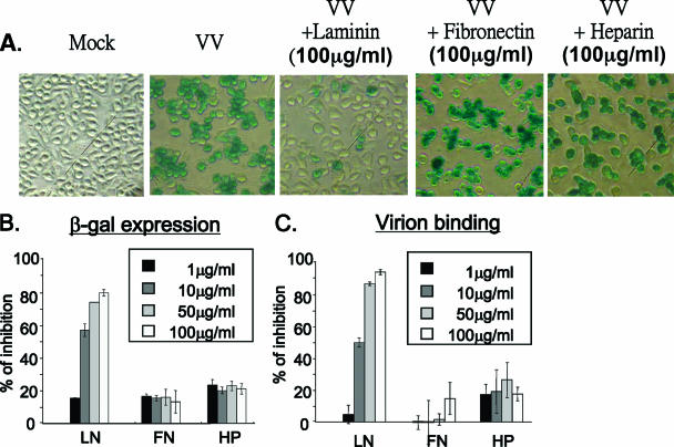
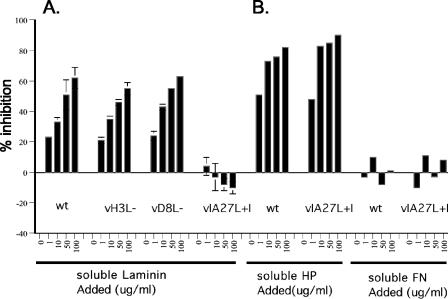
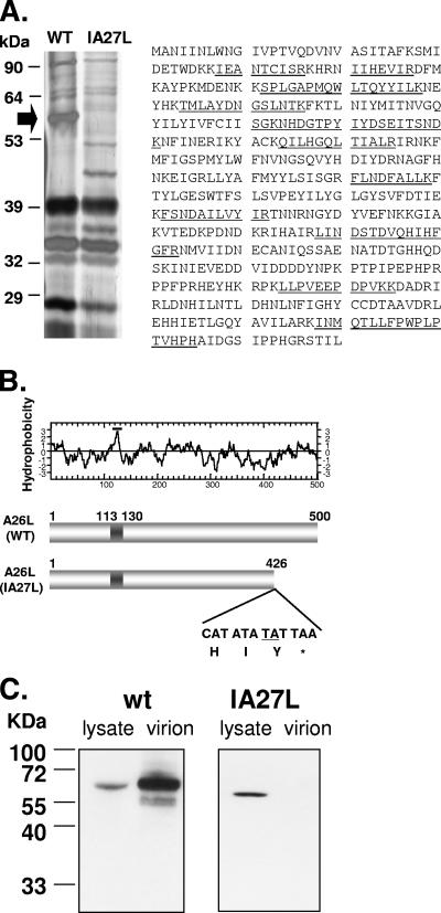
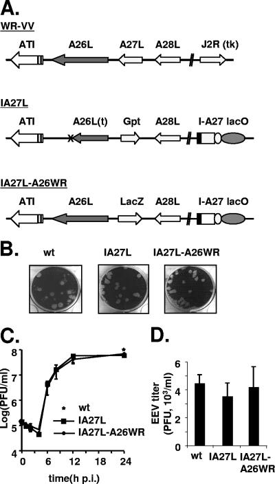
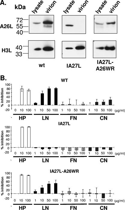
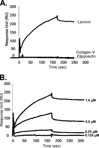
Vaccinia virus intracellular mature virus (IMV) binds to glycosaminoglycans (GAGs) on cells via three virion proteins, H3L, A27L, and D8L. In this study, we demonstrated that binding of IMV to BSC40 cells was competitively inhibited by soluble laminin but not by fibronectin or collagen V, suggesting that this cell surface extracellular matrix (ECM) protein may play a role in vaccinia virus entry. Moreover, IMV infection of GAG(-) sog9 cells was also inhibited by laminin, demonstrating that virion binding to laminin does not involve a prior interaction with GAGs. Furthermore, comparative envelope protein analyses of wild-type vaccinia virus strain Western Reserve, which binds to laminin, and of a mutant virus, IA27L, which does not, showed that the A26L open reading frame (ORF), encoding an envelope protein, was mutated in IA27L, resulting in A26L being absent from the IMV. Expression of the wild-type A26L ORF in IA27L resulted in laminin binding activity. Moreover, recombinant A26L protein bound to laminin in vitro with a high affinity, providing direct evidence that A26L is the laminin binding protein on IMV. In summary, these results reveal a novel role for the vaccinia viral envelope protein A26L in binding to the ECM protein laminin, an association that is proposed to facilitate IMV entry.


Figures

FIG. 1.
FIG. 1.
Laminin blocks IMV binding to BSC40 cells. (A) Different concentrations of soluble LN, FN, CN, or HP were mixed with purified IMV (150 PFU) for 30 min at 4°C and then added to cells for 30 min at 4°C, and the percent inhibition of virus binding to cells was determined by plaque assay as described in Materials and Methods. (B) Confocal immunofluorescence microscopy showing that soluble laminin reduces IMV binding to BSC40 cells. Cells were infected in the absence (VV) or presence (VV+LN) of 100 μg/ml of laminin at an MOI of 20 PFU per cell for 30 min at 4°C, and cell-bound virions were visualized by staining with anti-L1R antibody (red stain) and confocal microscopy as previously described (11, 48). The nucleus is stained blue with DAPI (4′,6′-diamidino-2-phenylindole).
FIG. 2.
FIG. 2.
Soluble laminin inhibits vaccinia virus infection of sog9 cells. (A) sog9 cells were infected with vMJ360 (expressing lacZ from a viral early promoter) at an MOI of 5 PFU per cell in the presence (100 μg/ml) or absence of soluble laminin, fibronectin, or heparin as described in the legend to Fig. 1. The infected cells were fixed at 3 h p.i. in 0.5% paraformaldehyde and analyzed for β-Gal activity by X-Gal staining. (B) Experiments were performed as for panel A except that different concentrations (0, 1, 10, 50, and 100 μg/ml) of soluble laminin, fibronectin, or heparin were used and β-Gal activity was quantified by an ONPG assay as described previously (18). (C) Experiments were performed as for panel B except that the cells were harvested and cell-bound IMV particles were determined by confocal immunofluorescence analysis as described in Materials and Methods.
FIG. 3.
FIG. 3.
Laminin blocks the binding of wild-type vaccinia virus IMV to BSC40 cells, but not that of IA27L. Purified IMVs from wild-type vaccinia virus (wt), H3L, D8L, or IA27L were mixed for 30 min at 4°C with different concentrations (0, 1, 10, 50, and 100 μg/ml) of soluble laminin (A) or HP or FN (B) and then were used to infect BSC40 cells, and the percent inhibition of virus binding to cells was determined as described in Materials and Methods.
FIG. 4.
FIG. 4.
A26L is absent in envelope proteins extracted from IA27L. (A) Silver staining of a 7% SDS-polyacrylamide gel showing IMV membrane proteins extracted from wild-type virus (WT) and IA27L using 1% NP-40 and 50 mM DTT (left panel). The arrow indicates the 58-kDa protein excised for protein identification by mass spectroscopy. Right panel: amino acid sequence of the A26L/WR149 protein taken from http://www.poxvirus.org/; the 11 tryptic peptides detected by mass spectrometry are underlined. (B) Upper panel: hydrophobicity plot of A26L from vaccinia virus Western Reserve strain. Region 113-130 contains hydrophobic residues and is a potential transmembrane region. Lower panel: insertion of nucleotides TA (underlined) in IA27L and the resulting premature termination codon TAA. WT, wild type. (C) Immunoblot analysis of A26L in lysates and purified IMVs from BSC40 cells infected with wild-type (wt) virus or IA27L using anti-A26L antibody.
FIG. 5.
FIG. 5.
Generation of recombinant IA27L-A26WR. (A) Schematic diagrams of wild-type vaccinia virus Western Reserve strain (WR-VV), IA27L, and IA27L expressing wild-type A26L (IA27L-A26WR). The A26L and J2R (tk) loci in the virus genomes are shown. In IA27L, the A27L locus is inactivated by Gpt insertion, and a gene cassette with an IPTG-inducible A27L ORF (I-A27) and the lacI repressor gene (lacO) is inserted into the J2R locus (35). The arrows indicate the direction of transcription. Expression of lacZ is driven by a viral late promoter, p11k. The X marks the TA insertion site in the A26L ORF in IA27L virus. (B) Plaque morphology of wild-type virus (wt), IA27L, and IA27L-A26WR after crystal violet staining. (C) IMV yield in one-step growth analyses of IA27L and IA27L-A26WR in BSC40 cells. BSC40 cells were infected with each virus at an MOI of 5 PFU per cell, incubated in culture medium with 5 mM IPTG, and harvested at the indicated time for plaque assays. The star shows the yield of wild-type virus (wt) at 24 h p.i. (D) EEV yield in one-step growth analyses of wild-type virus (wt), IA27L, and IA27L-A26WR. BSC40 cells were infected as described for panel C, the culture medium was collected at 24 h p.i. and mixed with anti-L1R antibody to block contaminating IMV and then was used to infect BSC40 cells, and the EEV titer was determined.
FIG. 6.
FIG. 6.
Expression of full-length A26L in IA27L-A26WR virus allows IMV binding to laminin. (A) Immunoblot analyses of A26L expression in cell lysates (24 h p.i.) and in purified virions from cells infected with wild-type virus (wt), IA27L, or IA27L-A26WR using anti-A26L or anti-H3L antibodies. (B) Soluble laminin blocks the binding of IA27L-A26WR to BSC40 cells. Soluble HP, LN, FN, and CN at the indicated concentrations were tested for their abilities to block the binding of wild-type (WT), IA27L, and IA27L-A26WR IMV virions to BSC40 cells, as described in the legend to Fig. 1.
FIG. 7.
FIG. 7.
(A) SPR analysis of the binding of A26L(127-500) to laminin, collagen V, or fibronectin. Biotinylated GST-A26L(127-500) was immobilized on an SA sensor flow chip and tested for the binding of 1 μM laminin, collagen V, or fibronectin as described in Materials and Methods; the first 180 s represent association, and the next 300 s represent dissociation. (B) SPR sensorgrams of laminin binding after removal of the background binding of laminin to biotinylated GST using four different concentrations of laminin (0.125, 0.25, 0.5, and 1.0 μM) with a flow rate of 10 μl/minute to extract the association/dissociation kinetics parameters. As a result, a kon of 1.5 × 10⁴ M−1 s−1, a koff of 2.0 × 10⁻⁴ s−1, and a KA (=kon/koff) of 7.5 × 10⁷ M−1 were determined for the A26L and laminin interaction (χ2, 9.6). The binding rate constants were derived from a 1:1 Langmuir binding model (1). The association/dissociation kinetics were measured and recorded by SPR as described in Materials and Methods.

References

    1. Adamson, A. W. 1967. Physical chemistry of surfaces, p. 397-401. Interscience, New York, NY.
    1. Armstrong, J. A., D. H. Metz, and M. R. Young. 1973. The mode of entry of vaccinia virus into L cells. J. Gen. Virol. 21:533-537. - PubMed
    1. Banfield, B. W., Y. Leduc, L. Esford, K. Schubert, and F. Tufaro. 1995. Sequential isolation of proteoglycan synthesis mutants by using herpes simplex virus as a selective agent: evidence for a proteoglycan-independent virus entry pathway. J. Virol. 69:3290-3298. - PMC - PubMed
    1. Barbosa, A. S., P. A. Abreu, F. O. Neves, M. V. Atzingen, M. M. Watanabe, M. L. Vieira, Z. M. Morais, S. A. Vasconcellos, and A. L. Nascimento. 2006. A newly identified leptospiral adhesin mediates attachment to laminin. Infect. Immun. 74:6356-6364. - PMC - PubMed
    1. Brown, E., T. G. Senkevich, and B. Moss. 2006. Vaccinia virus F9 virion membrane protein is required for entry but not virus assembly, in contrast to the related L1 protein. J. Virol. 80:9455-9464. - PMC - PubMed

Publication types

MeSH terms

LinkOut - more resources