Comparative Study
. 2006 Dec 13;26(50):13076-88.
doi: 10.1523/JNEUROSCI.3746-06.2006.

Progressive deafness and altered cochlear innervation in knock-out mice lacking prosaposin

Affiliations
Comparative Study

Progressive deafness and altered cochlear innervation in knock-out mice lacking prosaposin

Omar Akil et al. J Neurosci. .

Abstract

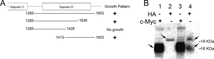
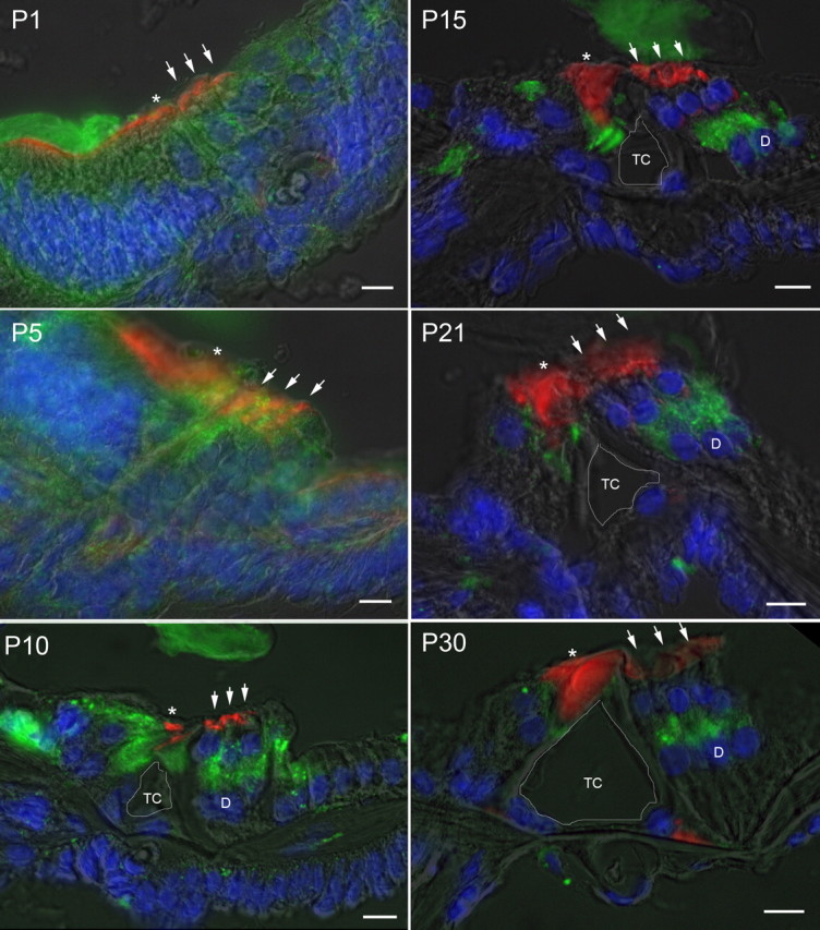
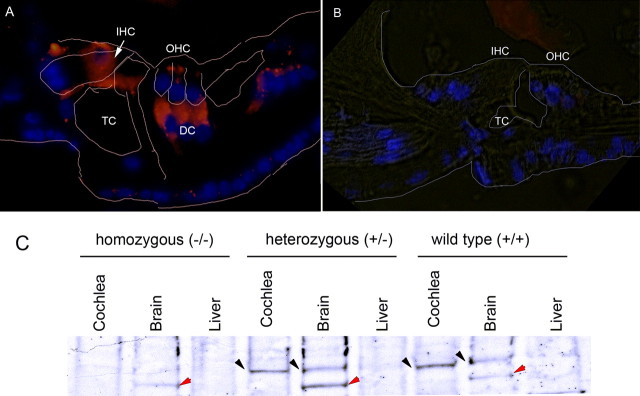
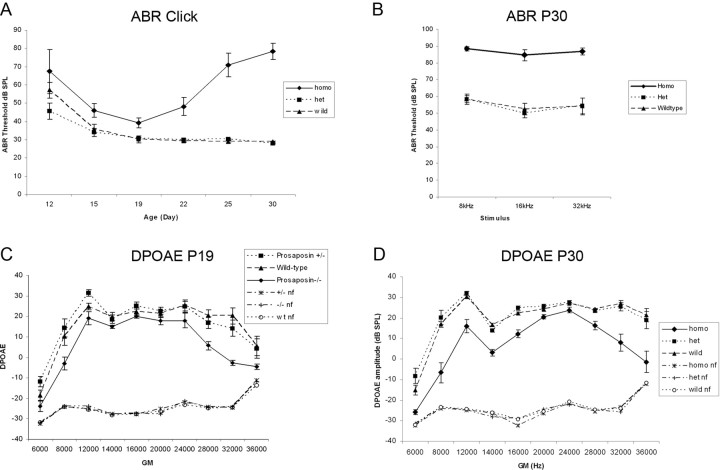
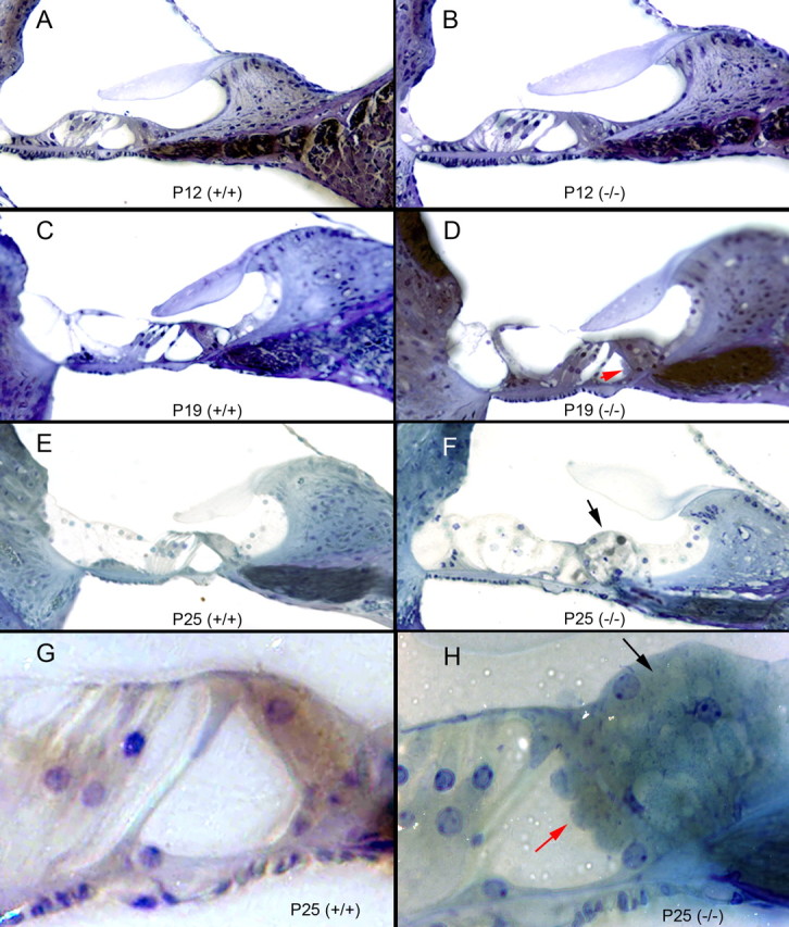
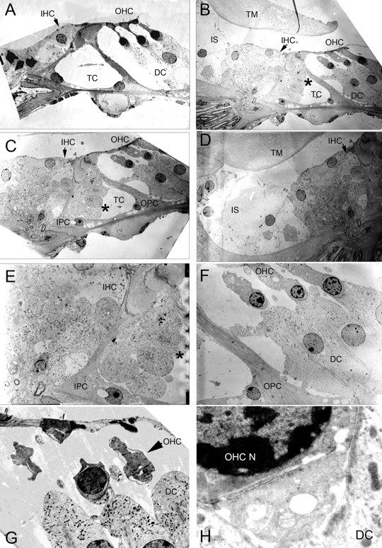
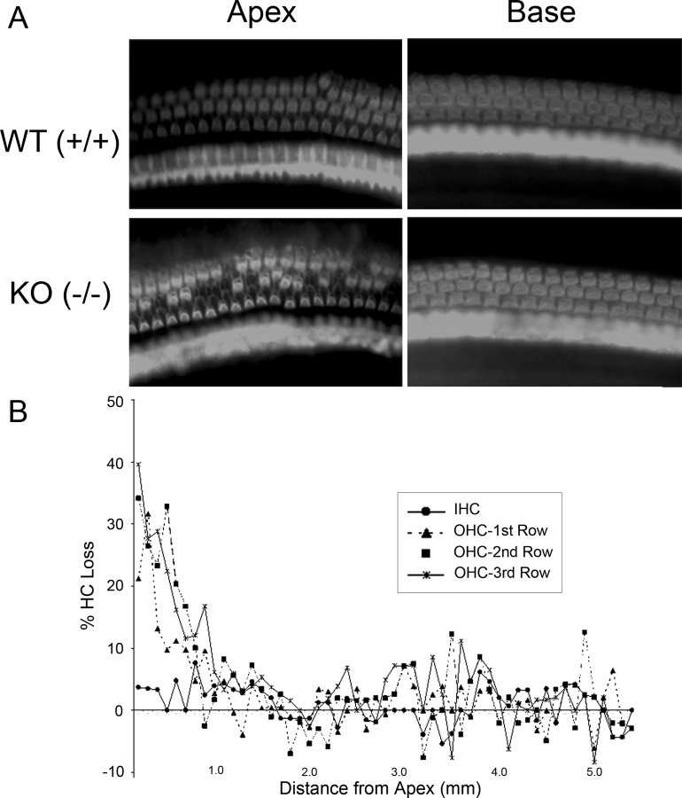
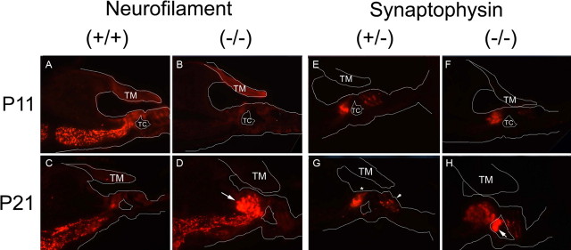
After a yeast two-hybrid screen identified prosaposin as a potential interacting protein with the nicotinic acetylcholine receptor (nAChR) subunit alpha10, studies were performed to characterize prosaposin in the normal rodent inner ear. Prosaposin demonstrates diffuse organ of Corti expression at birth, with gradual localization to the inner hair cells (IHCs) and its supporting cells, inner pillar cells, and synaptic region of the outer hair cells (OHCs) and Deiters' cells (DCs) by postnatal day 21 (P21). Microdissected OHC and DC quantitative reverse transcriptase-PCR and immunohistology localizes prosaposin mRNA to DCs and OHCs, and protein predominantly to the apex of the DCs. Subsequent studies in a prosaposin knock-out (KO) (-/-) mouse showed intact but slightly reduced hearing through P19, but deafness by P25 and reduced distortion product otoacoustic emissions from P15 onward. Beginning at P12, the prosaposin KO mice showed histologic organ of Corti changes including cellular hypertrophy in the region of the IHC and greater epithelial ridge, a loss of OHCs from cochlear apex, and vacuolization of OHCs. Immunofluorescence revealed exuberant overgrowth of auditory afferent neurites in the region of the IHCs and proliferation of auditory efferent neurites in the region of the tunnel of Corti. IHC recordings from these KO mice showed normal I-V curves and responses to applied acetylcholine. Together, these results suggest that prosaposin helps maintain normal innervation patterns to the organ of Corti. Furthermore, prosaposin's overlapping developmental expression pattern and binding capacity toward the nAChR alpha10 suggest that alpha10 may also play a role in this function.


Figures

Figure 1.
Figure 1.
Verification of the yeast two-hybrid interaction. A, Varying lengths of the terminal end of prosaposin (corresponding to saposin D) were used in yeast two-hybrid constructs to test the protein–protein interaction. The region that appears to be primarily responsible for the interaction with nAChR α10 resides between 1428 and 1536 of saposin D. B, In vitro co-IP of nAChR α10 and prosaposin. Lane 1 is α10 alone labeled by c-Myc (arrow next to strongly labeled band), probing with a c-Myc antibody. Lane 2 is prosaposin alone labeled by hemagglutinin (HA; arrow), probing with an HA antibody. Lane 3 is a co-IP labeling α10 (strongly) and prosaposin (weakly) probing with the c-Myc antibody. As expected, both bands corresponding to prosaposin and α10 are present. The larger additional bands noted are also seen in lane 1 and represent nonspecific binding of c-myc/α10. Lane 8 is a co-IP labeling both α10 and prosaposin (arrows) probing with the HA antibody. This experiment independently verifies protein–protein binding between the intracellular loop of nAChR α10 and prosaposin.
Figure 2.
Figure 2.
Developmental expression of prosaposin in rat cochlea. Immunofluorescence staining of prosaposin (green), rhodamine-phalloidin (red), and DAPI (blue) with a semitranslucent transmitted light micrograph overlay from P1 through P30 demonstrates initially diffuse expression of prosaposin throughout the primitive organ of Corti that gradually localizes to several discrete locations within the organ of Corti, including the IHCs and its supporting cells, inner pillar cells, and synaptic region of the OHCs (either base of OHCs or apex of the DCs). At P5 there is increased signal in the regions of the IHC and OHC relative to surrounding tissues. By P10, labeling can be clearly differentiated in the region of the basal OHCs or apical DCs, IHCs, and inner pillar cells. This labeling pattern is refined, with further loss of label in surrounding cells through P30, with localization to regions surrounding the IHC, inner pillar cell, and base of OHC and/or apex of DC. In each panel, an asterisk is placed above the IHC, three small arrows are placed above the OHC, and the central of the three Deiters' cells is marked by a D where clearly visible (after P10). The tunnel of Corti is outlined in white and labeled TC. Scale bars, 10 μm.
Figure 3.
Figure 3.
Localization of prosaposin in the organ of Corti by RT-PCR and immunohistology. Multiple modalities were used to localize prosaposin in the region of the OHCs and DCs. A, RT-PCR from microdissected rat OHCs. A first round RT-PCR (left) demonstrates the presence of α9 (arrow; used here as an OHC marker; similar results were obtained with α10) (data not shown) from OHCs, but not DCs. Similarly, a whole section of the organ of Corti (OC) demonstrates a strong band of α9. To verify that DCs have not contaminated the OHC sample, the presence of Glast1 mRNA (arrow; present only in DCs) is tested using two successive rounds of PCR (middle). The right panel demonstrates the presence of both α9 and prosaposin (Psap) in OHCs, but not in the background fluid (Fl), clearly indicating the presence of prosaposin mRNA in OHCs. B, Double labeling of rat organ of Corti with prosaposin (red) and phalloidin (Phal, green) demonstrates that prosaposin lies predominantly below the basal region of the OHCs (arrow, outline of OHC base), in the efferent synaptic cleft and/or Deiters' cell. Intense prosaposin labeling is also noted in the IHC region (asterisk). C, Double labeling of rat organ of Corti with prosaposin (red) and synaptophysin (green) demonstrates that prosaposin predominantly lies at or below the efferent synaptic cleft (arrow) below the base of the OHC, again suggesting predominant prosaposin labeling in the apex of the DCs or efferent synaptic cleft. Once again, strong prosaposin labeling is also noted in the IHC (asterisk). TM, Tectorial membrane. D, E, Triple labeling of prosaposin (red), ChAT (green), and DAPI (blue labels nuclei) in either rat (D) or mouse (E) organ of Corti, demonstrates that prosaposin resides largely in the efferent synaptic cleft and the apex of the DCs. For orientation, the OHCs, pillar cells, and IHC (asterisk) are outlined in white, whereas the tunnel of Corti (TC) is labeled, as are the third OHC and DC nuclei. F, To resolve the discrepancy of the RT-PCR and immunofluorescence results regarding prosaposin in OHCs, quantitative real-time PCR of microdissected OHCs and DCs was performed. Compared with L19, a ribosomal housekeeping gene, prosaposin shows a 4-fold amplification in OHCs (P-OHC) and a 6.5-fold amplification in DCs (P-DC), indicating higher levels of prosaposin mRNA in DCs as compared with OHCs. For comparison, both nAChR α9 and α10 show a marked decrease in concentration as compared with L19 in OHCs (α9-OHC and α10-OHC, respectively). As a control, there is no α10 identified in DCs, indicating a relatively pure population of cells. Together, these studies suggest that prosaposin exists mostly in DCs or the efferent synaptic cleft, whereas the OHCs contains a much smaller concentration of prosaposin. Error bars indicate SEM. Scale bars, 10 μm.
Figure 4.
Figure 4.
Prosaposin expression in the null mutant (knock-out) mouse. Prosaposin antibody labeling demonstrates the absence of prosaposin in the KO mouse organ of Corti. A, B, Compared with WT mice (A), homozygote prosaposin KO mice lack prosaposin expression within the organ of Corti (B). TC, Tunnel of Corti. C, In the homozygous (KO) mice, Western blot analysis demonstrates a lack of prosaposin expression within the brain, a known location of prosaposin, in contrast to heterozygote and WT mice (black arrow). In all three brain lanes, there is nonspecific banding (red arrows); because the band also appears in the KO sample, it is not prosaposin labeling. The nonspecific band does not appear in the cochlear samples, and, thus, the labeling seen in the cochlea likely represents true prosaposin labeling. The brain serves as a positive control whereas the liver serves as a negative control in each experiment.
Figure 5.
Figure 5.
Hearing in the prosaposin knock-out mice. A, B, Mice at varying ages were tested through P30 (A) using clicks, and at 8, 16, and 32 kHz (B). A, All mice demonstrate normal auditory development through P19, although ABR thresholds are statistically significantly higher in the homozygotes as compared with heterozygotes and WT. After P19, however, hearing in the homozygote KO mice declines rapidly whereas hearing stays normal in the heterozygote and WT mice. C, DPOAEs were obtained from mice at these same ages. Before P19, DPOAE amplitudes are significantly reduced at very low (8 kHz) and high (32 kHz) frequencies for the homozygotes as compared with heterzygotes and WT mice. D, By P30, DPOAEs are significantly reduced at all frequencies tested for the KO mice as compared with heterozygotes and WT. nf, Noise floor.
Figure 6.
Figure 6.
Cochlear histology in the prosaposin knock-out mice; mouse organ of Corti histology when the neuritogenic region of saposin C is mutated to become nonfunctional. A, C, E, G, Normal P12, P19, and P25 (low and high magnification) wild-type (+/+) mouse organ of Corti histology (A, C, E, 40× magnification; G, 100× magnification). B, D, F, H, The homozygous (−/−) mouse organ of Corti at varying ages (P12, P19, and P25 at low and high magnification). The gross morphology (e.g., number of turns) is identical to the wild-type, as is the spiral ganglion cell number and cellular morphology. Beginning at P15 and increasing through P30, there is marked hyperplasia of cells surrounding the inner hair cell and in the region of the inner sulcus and greater epithelial (F, H, black arrow highlights this change). There is also some tissue bulging into the tunnel of Corti from the inner hair cell region, noted as early as P15, and here labeled at P19 and P25 (D, H, red arrows). DCs also appear larger than normal with an ill-defined border between the DCs and the OHCs in animals beyond P25 (most notable in F).
Figure 7.
Figure 7.
Cochlear electron microscopy of the prosaposin knock-out mice. A, Wild-type organ of Corti demonstrating normal anatomy. B, C, In the prosaposin KO mouse, there is marked cellular hypertrophy in the region of the IHCs. B, C, E, There is bulging tissue into the tunnel of Corti (TC, asterisk). D, Hypertrophy of the inner sulcus (IS) region. FH, In the region of the OHCs, DCs are hypertrophied and there is vacuolization within the OHCs. H, The base of the OHC, efferent synaptic cleft, and apex of the DC. The synaptic cleft between the OHC and the DCs is abnormally enlarged and vacuolization is seen in the cells. TM, Tectorial membrane; OPC, outer pillar cell.
Figure 8.
Figure 8.
Hair cell counts in the prosaposin knock-out mice. A, Representative samples of phalloidin-stained cochlear preparations in the base and apex of WT and KO mice. There is a loss of OHCs in the apex of the KO mice. B, Hair cell cytocochleogram in the organ of Corti. Compared with WT mice, there is a loss of up to 40% of OHCs in the cochlear apex of the prosaposin KO mice, extending through the first 2 mm of the cochlear duct. In contrast, IHC numbers are normal throughout the cochlea.
Figure 9.
Figure 9.
Cochlear immunohistochemistry of the prosaposin knock-out mice. Immunofluorescent labeling with neurofilament and synaptophysin antibodies in the knock-out mice organ of Corti lacking a functional neuritogenic region of saposin C. A white outline of relevant structures within the organ of Corti are added within each picture to orient the staining pattern. AD, Neurofilament-200 (afferent auditory neuronal) staining at both P11 (A, B) and P21 (C, D). The staining pattern shows intense labeling at P11 in the wild-type (identical to the heterozygote) (data not shown), but weak labeling in the homozygote by comparison. By P21, the heterozygote has a normal amount of labeling, whereas the homozygote has an abundance of labeling in the region corresponding the hypercellular region surrounding the inner hair cell seen in Figures 6 and 7. EH, Synaptophysin (efferent auditory neuronal) staining at P11 (E, F) and P21 (G, H). At P11, there is very little difference between wild-type (data not shown), heterozygous (E) and homozygous (F) knock-out mice organ of Corti. By P21, in the WT (data not shown) and heterozygote (G), staining is noted in the region of the IHC (below asterisk) and base of the OHCs (small arrow), as expected. In contrast, in the KO mice (H), there is a significant amount of synaptophysin staining in the regions of the tunnel of Corti (TC; arrow) and to a lesser extent, the region surrounding the IHC. There is limited staining noted in the region of the OHCs in both P11 and P21 homozygotes as compared with heterozygotes or WT at both ages. The cellular bulging into the tunnel of Corti seen in Figure 6 and 7 corresponds exactly to the intense synaptophysin staining here in the homozygote (H). TM, Tectorial membrane.
Figure 10.
Figure 10.
Hair cell physiology in the prosaposin knock-out mice. Cholinergic efferent synapses at the IHC show normal function in prosaposin (−/−) mice. Animals were tested before (P7–P11) and after (P19) the onset of hearing. A, B, At P7–P11, IHC IV relationships showed Ca2+ inward currents and delayed rectifier potassium currents. At P19, IHC IV relationships also exhibited substantial BK potassium currents, which can be identified by a fast activation time compared with the slower activation time of delayed rectifier potassium currents. C, E, At age P7–P11, prosaposin (−/−) mice, like the wild-type, exhibited ACh-activated currents and efferent synaptic currents in IHCs. The ACh-activated (100 μm) currents were inward at a holding potential of −90 mV and outward at −50 mV, suggesting that both ACh receptors and SK current were functional. Application of 25 mm potassium induced an inward current in IHCs, and also activated synaptic activity. D, F, At age P19, prosaposin (−/−) mice showed a phenotype as expected for a normal development for efferent innervation: application of ACh did not elicit any currents. Application of 25 (or 40) mm potassium induced an inward current caused by the change in the potassium reversal potential but did not activate any efferent synaptic activity.

References

    1. Baker ER, Zwart R, Sher E, Millar NS. Pharmacological properties of α9α10 nicotinic acetylcholine receptors revealed by heterologous expression of subunit chimeras. Mol Pharmacol. 2004;65:453–460. - PubMed
    1. Berglund AM, Ryugo DK. Neurofilament antibodies and spiral ganglion neurons of the mammalian cochlea. J Comp Neurol. 1991;306:393–408. - PubMed
    1. Campana WM, Eskeland N, Calcutt NA, Misasi R, Myers RR, O'Brien JS. Prosaptide prevents paclitaxel neurotoxicity. Neurotoxicology. 1998;19:237–244. - PubMed
    1. Campana WM, Mohiuddin L, Misasi R, O'Brien JS, Calcutt NA. Prosaposin-derived peptides enhanced sprouting of sensory neurons in vitro and induced sprouting at motor endplates in vivo. J Peripher Nerv Syst. 2000;5:126–130. - PubMed
    1. Elgoyhen AB, Johnson DS, Boulter J, Vetter DE, Heinemann S. α9: an acetylcholine receptor with novel pharmacological properties expressed in rat cochlear hair cells. Cell. 1994;79:705–715. - PubMed

Publication types

MeSH terms