

Role of Kupffer cells in the pathogenesis of liver disease
- PMID: 17167827
- PMCID: PMC4087584
- DOI: 10.3748/wjg.v12.i46.7413
Role of Kupffer cells in the pathogenesis of liver disease
Abstract
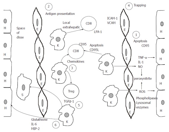
Kupffer cells, the resident liver macrophages have long been considered as mostly scavenger cells responsible for removing particulate material from the portal circulation. However, evidence derived mostly from animal models, indicates that Kupffer cells may be implicated in the pathogenesis of various liver diseases including viral hepatitis, steatohepatitis, alcoholic liver disease, intrahepatic cholostasis, activation or rejection of the liver during liver transplantation and liver fibrosis. There is accumulating evidence, reviewed in this paper, suggesting that Kupffer cells may act both as effector cells in the destruction of hepatocytes by producing harmful soluble mediators as well as antigen presenting cells during viral infections of the liver. Moreover they may represent a significant source of chemoattractant molecules for cytotoxic CD8 and regulatory T cells. Their role in fibrosis is well established as they are one of the main sources of TGFbeta1 production, which leads to the transformation of stellate cells into myofibroblasts. Whether all these variable functions in the liver are mediated by different Kupffer cell subpopulations remains to be evaluated. In this review we propose a model that demonstrates the role of Kupffer cells in the pathogenesis of liver disease.
Figures
References
-
- Bouwens L, Baekeland M, De Zanger R, Wisse E. Quantitation, tissue distribution and proliferation kinetics of Kupffer cells in normal rat liver. Hepatology. 1986;6:718–722. - PubMed
-
- MacPhee PJ, Schmidt EE, Groom AC. Evidence for Kupffer cell migration along liver sinusoids, from high-resolution in vivo microscopy. Am J Physiol. 1992;263:G17–G23. - PubMed
-
- Nolan JP. Endotoxin, reticuloendothelial function, and liver injury. Hepatology. 1981;1:458–465. - PubMed
-
- Bayón LG, Izquierdo MA, Sirovich I, van Rooijen N, Beelen RH, Meijer S. Role of Kupffer cells in arresting circulating tumor cells and controlling metastatic growth in the liver. Hepatology. 1996;23:1224–1231. - PubMed
Publication types
MeSH terms
LinkOut - more resources
Full Text Sources
Other Literature Sources
Medical
Research Materials
