

Apoptosis in skeletal muscle and its relevance to atrophy
- PMID: 17167834
- PMCID: PMC4087591
- DOI: 10.3748/wjg.v12.i46.7463
Apoptosis in skeletal muscle and its relevance to atrophy
Abstract
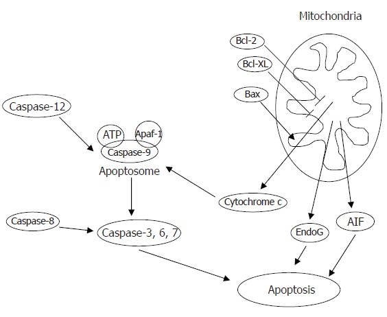
Apoptosis is necessary for maintaining the integrity of proliferative tissues, such as epithelial cells of the gastrointestinal system. The role of apoptosis in post mitotic tissues, such as skeletal muscle, is less well defined. Apoptosis during muscle atrophy occurs in both myonuclei and other muscle cell types. Apoptosis of myonuclei likely contributes to the loss of muscle mass, but the mechanisms underlying this process are largely unknown. Caspase-dependent as well as -independent pathways have been implicated and the mode by which atrophy is induced likely determines the apoptotic mechanisms that are utilized. It remains to be determined whether a decrease in apoptosis will alleviate atrophy and distinct research strategies may be required for different causes of skeletal muscle loss.
Figures
Similar articles
-
[Apoptosis in atrophic skeletal muscle induced by brachial plexus injury in rats].Zhonghua Yi Xue Za Zhi. 2000 Jul;80(7):530-3. Zhonghua Yi Xue Za Zhi. 2000. PMID: 11798813 Chinese.
-
Apoptosis in the skeletal muscle of patients with heart failure: investigation of clinical and biochemical changes.Heart. 2000 Oct;84(4):431-7. doi: 10.1136/heart.84.4.431. Heart. 2000. PMID: 10995417 Free PMC article.
-
Mitochondrial Apoptotic Signaling Involvement in Remodeling During Myogenesis and Skeletal Muscle Atrophy.Semin Cell Dev Biol. 2023 Jul 15;143:66-74. doi: 10.1016/j.semcdb.2022.01.011. Epub 2022 Feb 28. Semin Cell Dev Biol. 2023. PMID: 35241367 Review.
-
Age-related differences in apoptosis with disuse atrophy in soleus muscle.Am J Physiol Regul Integr Comp Physiol. 2005 May;288(5):R1288-96. doi: 10.1152/ajpregu.00576.2004. Epub 2005 Jan 13. Am J Physiol Regul Integr Comp Physiol. 2005. PMID: 15650125
-
Apoptosis in muscle atrophy: relevance to sarcopenia.Exp Gerontol. 2005 Jun;40(6):473-81. doi: 10.1016/j.exger.2005.04.003. Exp Gerontol. 2005. PMID: 15935591 Review.
Cited by
-
The senescent rat diaphragm does not exhibit age-related changes in caspase activities, DNA fragmentation, or myonuclear domain.Eur J Appl Physiol. 2012 Dec;112(12):3983-90. doi: 10.1007/s00421-012-2380-2. Epub 2012 Mar 21. Eur J Appl Physiol. 2012. PMID: 22434253
-
Further considerations on in vitro skeletal muscle cell death.Muscles Ligaments Tendons J. 2014 Feb 24;3(4):267-74. eCollection 2013 Oct. Muscles Ligaments Tendons J. 2014. PMID: 24596689 Free PMC article.
-
Enhanced survival of skeletal muscle myoblasts in response to overexpression of cold shock protein RBM3.Am J Physiol Cell Physiol. 2011 Aug;301(2):C392-402. doi: 10.1152/ajpcell.00098.2011. Epub 2011 May 18. Am J Physiol Cell Physiol. 2011. PMID: 21593448 Free PMC article.
-
Chronic Binge Alcohol-Induced Dysregulation of Mitochondrial-Related Genes in Skeletal Muscle of Simian Immunodeficiency Virus-Infected Rhesus Macaques at End-Stage Disease.Alcohol Alcohol. 2017 May 1;52(3):298-304. doi: 10.1093/alcalc/agw107. Alcohol Alcohol. 2017. PMID: 28069597 Free PMC article.
-
Taurine Attenuates Disuse Muscle Atrophy Through Modulation of the xCT-GSH-GPX4 and AMPK-ACC-ACSL4 Pathways.Antioxidants (Basel). 2025 Jul 10;14(7):847. doi: 10.3390/antiox14070847. Antioxidants (Basel). 2025. PMID: 40722950 Free PMC article.
References
-
- Primeau AJ, Adhihetty PJ, Hood DA. Apoptosis in heart and skeletal muscle. Can J Appl Physiol. 2002;27:349–395. - PubMed
-
- Earnshaw WC, Martins LM, Kaufmann SH. Mammalian caspases: structure, activation, substrates, and functions during apoptosis. Annu Rev Biochem. 1999;68:383–424. - PubMed
-
- Candé C, Vahsen N, Garrido C, Kroemer G. Apoptosis-inducing factor (AIF): caspase-independent after all. Cell Death Differ. 2004;11:591–595. - PubMed
-
- Li LY, Luo X, Wang X. Endonuclease G is an apoptotic DNase when released from mitochondria. Nature. 2001;412:95–99. - PubMed
-
- van Loo G, Schotte P, van Gurp M, Demol H, Hoorelbeke B, Gevaert K, Rodriguez I, Ruiz-Carrillo A, Vandekerckhove J, Declercq W, et al. Endonuclease G: a mitochondrial protein released in apoptosis and involved in caspase-independent DNA degradation. Cell Death Differ. 2001;8:1136–1142. - PubMed
Publication types
MeSH terms
Substances
Grants and funding
LinkOut - more resources
Full Text Sources
Other Literature Sources
