

Review
doi: 10.1186/ar2075.
What is MRI bone oedema in rheumatoid arthritis and why does it matter?
Affiliations
- PMID: 17169137
- PMCID: PMC1794510
- DOI: 10.1186/ar2075
Item in Clipboard
Review
What is MRI bone oedema in rheumatoid arthritis and why does it matter?
Arthritis Res Ther.
2006.
Abstract
MRI bone oedema occurs in various forms of inflammatory and non-inflammatory arthritis and probably represents a cellular infiltrate within bone. It is common in early rheumatoid arthritis and is associated with erosive progression and poor functional outcome. Histopathological studies suggest that a cellular infiltrate comprising lymphocytes and osteoclasts may be detected in subchondral bone and could mediate the development of erosions from the marrow towards the joint surface. There is emerging evidence from animal models that such an infiltrate corresponds with MRI bone oedema, pointing towards the bone marrow as a site for important pathology driving joint damage in rheumatoid arthritis.
Figures
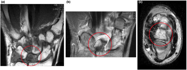
MRI scans from a 65 year old female rheumatoid arthritis patient with disease duration of one year. (a) Coronal T1 weighted image of the dominant wrist with reduced signal indicating florid bone oedema involving the entire lunate bone (circle). (b) Equivalent image following the injection of contrast (gadolinium diethylenetriamine pentaacetic acid (GdDPTA)) shows very bright signal within the lunate, suggesting the presence of vascularized tissue (slice does not exactly correspond with pre-GdDPTA image). (c) Axial T2w image with bright signal confirming bone oedema at the lunate.

Potential role of bone marrow-derived stem cells in trafficking to the subchondral bone and synovial membrane in rheumatoid arthritis joints, resulting in a subchondral cellular infiltrate (seen as bone oedema on MRI) followed by erosion. (a) CD34+ stem cells from bone marrow express high levels of NFkB, which leads to unusual sensitivity to TNFα. (b) Stem cells differentiate into fibroblast-like cells and travel via the circulation to synovial membrane to become type B synoviocytes – here they mediate formation of erosions via production of proinflammatory cytokines and matrix metalloproteinases [23,25]. (c) Stem cells may also traffic to the subchondral bone marrow where they differentiate into mesenchymal cells. These cells could then travel via bony canals from bone marrow to synovium [29] to excite an inflammatory response. (d) Alternatively, stem cells could travel to subchondral bone and at this site could mediate an inflammatory response via T/B cell interactions associated with angiogenesis [26] and osteoclast activation. This could lead to erosions originating from inside the bone, directed outwards towards joint surface [14]. (e) Coronal T2 weighted MRI scan of the wrist in early rheumatoid arthritis reveals bone oedema at the bases of the 2nd and 3rd metacarpals and adjacent regions of trapezoid and capitate carpal bones. Small intraosseous erosions are also apparent.
References
-
- The Collected Letters of Antoni van Leeuwenhoek 1701–1704. Vol. 14. ISBN: 9026514506. Lisse, The Netherlands: Swets and Zeitlinger; 1996.
-
- Lecouvet FE, van de Berg BC, Maldague BE, Lebon CJ, Jamart J, Saleh M, Noel H, Malghem J. Early irreversible osteonecrosis versus transient lesions of the femoral condyles: prognostic value of subchondral bone and marrow changes on MR imaging. AJR. 1998;170:71–77. - PubMed
Publication types
MeSH terms
LinkOut - more resources
Full Text Sources
Medical
