. 2007 Jan 26;1130(1):17-30.
doi: 10.1016/j.brainres.2006.10.083. Epub 2006 Dec 12.

Tight junction protein expression and barrier properties of immortalized mouse brain microvessel endothelial cells

Affiliations

Tight junction protein expression and barrier properties of immortalized mouse brain microvessel endothelial cells

Rachel C Brown et al. Brain Res. .

Abstract

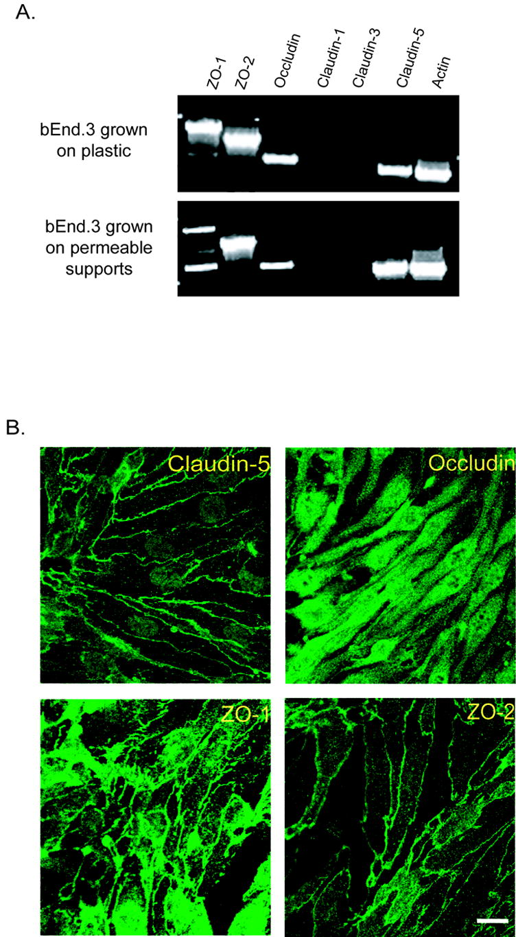
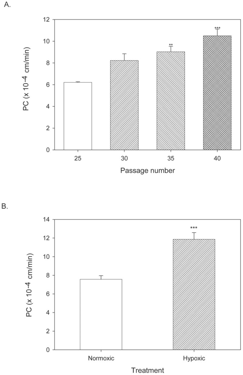
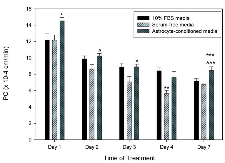
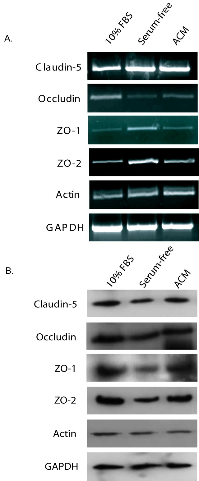
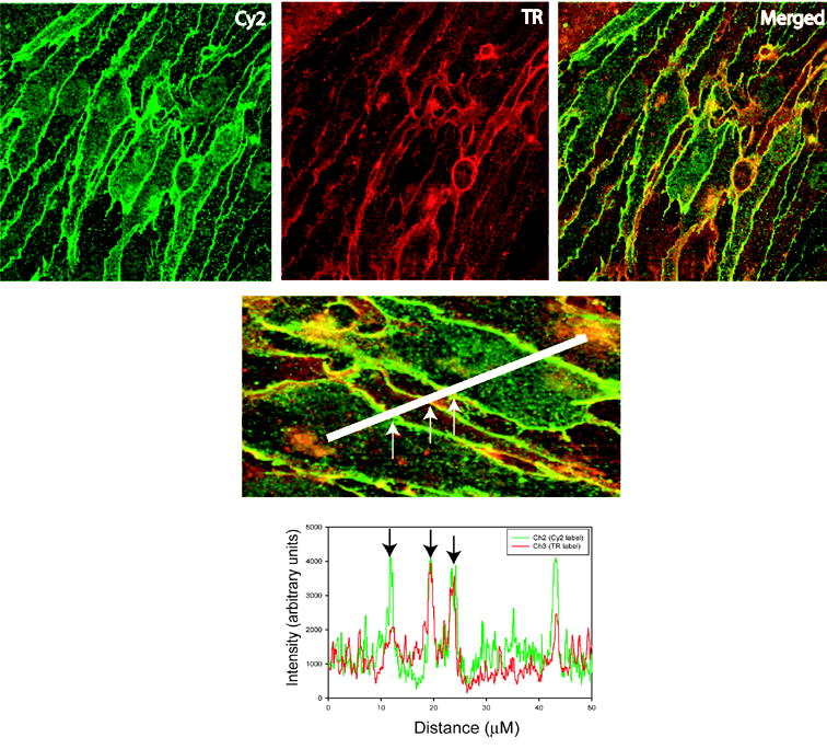
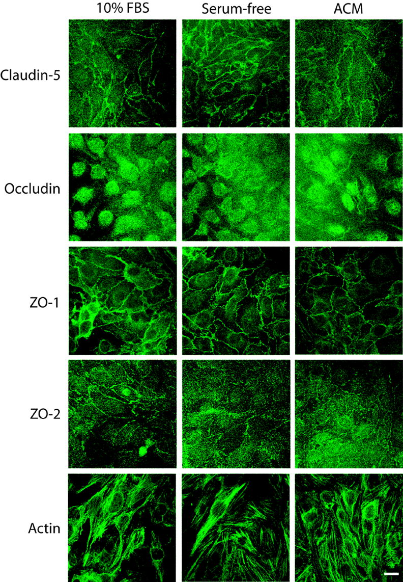
Understanding the molecular and biochemical mechanisms regulating the blood-brain barrier is aided by in vitro model systems. Many studies have used primary cultures of brain microvessel endothelial cells for this purpose. However, primary cultures limit the generation of material for molecular and biochemical assays since cells grow slowly, are prone to contamination by other neurovascular unit cells, and lose blood-brain barrier characteristics when passaged. To address these issues, immortalized cell lines have been generated. In these studies, we assessed the suitability of the immortalized mouse brain endothelial cell line, bEnd3, as a blood-brain barrier model. RT-PCR and immunofluorescence indicated expression of multiple tight junction proteins. bEnd3 cells formed barriers to radiolabeled sucrose, and responded like primary cultures to disrupting stimuli. Exposing cells to serum-free media on their basolateral side significantly decreased paracellular permeability; astrocyte-conditioned media did not enhance barrier properties. The serum-free media-induced decrease in permeability was correlated with an increase in claudin-5 and zonula occludens-1 immunofluorescence at cell-cell contracts. We conclude that bEnd3 cells are an attractive candidate as a model of the blood-brain barrier due to their rapid growth, maintenance of blood-brain barrier characteristics over repeated passages, formation of functional barriers and amenability to numerous molecular interventions.


Figures

FIGURE 1
FIGURE 1
Expression of tight junction protein mRNA and protein in bEnd3 cells. A) RNA was isolated from confluent bEnd3 cells grown on plastic cell culture dishes or on permeable Transwell™ filters, and RT-PCR was performed for ZO-1, ZO-2, occludin, claudin-1, claudin-3, claudin-5 and actin. bEnd3 cells express mRNA for ZO-1, ZO-2, occludin, claudin-5 and actin, but not for claudin-1 or claudin-3. This pattern of mRNA expression is not altered by the surface the cells are grown on. B) Expression of tight junction protein in bEnd3 cells grown on solid supports was assessed using confocal microscopy as described in the Materials and Methods. bEnd3 cells express immunoreactivity for claudin-5, occludin, ZO-1 and ZO-2. With the exception of occludin immunofluorescence, these proteins are localized to the cell membrane, as anticipated, where they are presumably part of the tight junction complex. Scale bar = 20 μm.
FIGURE 2
FIGURE 2
Permeability of bEnd3 monolayers with increasing passage number and in response to a disruptive stimulus. A) bEnd3 cells were passaged, and basal permeability (with 10% FBS in basolateral chamber) was assessed every five passages from P25 to P40. Cells were grown to confluency (6–7 days post-seeding) and permeability measurements were done 3–5 days later. There was a significant increase in basal permeability to [14C]sucrose at P35 and P40 as compared to P25. All further studies were performed before P35. ** p<0.01 vs. P25, *** p<0.001 vs. P25. n=6–8 filters. B) Hypoxic stress is a well known disruptor of the BBB [24,32]. bEnd3 monolayers were grown to confluence and exposed to 6 hr of hypoxic stress by incubation in 1% O2 as previously described [24,85]. Hypoxia increased bEnd3 monolayer permeability significantly, as seen in past studies with primary cultures [24]. n=4 filters, *** p<0.001 vs. control.
FIGURE 3
FIGURE 3
bEnd3 monolayer permeability is modulated by basolateral culturing conditions. bEnd3 cells were grown to confluence, and the media in the basolateral chamber was changed to either 10% FBS containing media, serum-free media, or a 50:50 mixture of serum-free media and astrocyte-conditioned media. After 1–7 days exposure to different media conditions, paracellular permeability of [14C]sucrose was measured. Permeability of the monolayers decreased over time in all treatment groups. For the first three days of exposure, there was a significant loosening of the monolayers exposed to astrocyte-conditioned media as compared to control (*) or serum-free media (^). By day 4, the monolayers exposed to serum-free media were significantly tighter then those exposed to 10% FBS or astrocyte-conditioned media. By day 7, there was no significant difference between 10% FBS and serum-free media exposed monolayers, while the monolayers exposed to astrocyte-conditioned media were again more permeable then the other two groups. n=8–16 filters, * p<0.05 versus 10% FBS, ** p<0.01 versus 10% FBS, *** p<0.001 versus 10% FBS, ^ p<0.05 versus serum-free media, ^^^ p<0.001 versus serum-free media.
FIGURE 4
FIGURE 4
Expression of tight junction mRNA and protein in bEnd3 cells after four days of exposure to 10% FBS, serum-free or astrocyte conditioned media. A) Confluent bEnd3 monolayers on permeable filters were exposed to the three types of media for four days, RNA was isolated, and RT-PCR was performed for tight junction proteins and for GAPDH as a loading control. Expression of all tight junction proteins was normalized to GAPDH expression to account for variations in starting material. There was no change in mRNA for claudin-5 or actin with treatment. Both ZO-1 and ZO-2 showed a slight increase in message in monolayers exposed to serum-free media for four days. Occludin message levels were decreased in monolayers exposed to serum-free or astrocyte-conditioned media. Representative blots are shown, n=3 experiments. B) Protein levels of tight junction components were assessed by immunoblotting with normalization to GAPDH band intensity as a loading control. There is a slight decrease in the levels of ZO-1 protein in monolayers exposed to serum-free media for four days. There are no significant changes in any of the other proteins examined. Representative blots are shown, n=3–4 experiments.
FIGURE 5
FIGURE 5
Analysis of confocal microscopy images. Representative confocal image is shown. bEnd3 cells were stained with antibody to claudin-5 (Cy2 panel) and wheat germ agglutinin (TR panel). The merged panel shows the overlap between claudin-5 immunoreactivity and wheat germ agglutinin incorporation in yellow. Lower panel shows how cells were sampled using a linescan measurement and generation of intensity histograms. Immunofluorescent intensities for membrane localizations were taken by averaging 2–5 μm of the linescan where overlap of the Cy2 and TR signals occurred, as shown in the histogram (arrows). Cytoplasmic intensities were taken from 2–5 μm regions with lowest TR signal, indicating the interior of the cell.
FIGURE 6
FIGURE 6
Subcellular localization of tight junction proteins after 4 days exposure to different basolateral media conditions. bEnd3 cells were grown to confluence on permeable filters and exposed to growth media (10% FBS), serum-free media (serum-free) or astrocyte-conditioned media (ACM) for four days. Monolayers were fixed and stained as described, and confocal images were collected. Images were analyzed by profiling fluorescence intensity at the cell membrane versus the cytoplasm; cell membranes were identified by co-staining with rhodamine-tagged wheat germ agglutinin (data not shown). The only proteins to show a significant alteration in subcellular localization with different treatments were claudin-5 and ZO-1, which shifted to a more-membrane localized position when exposed to either serum-free or astrocyte-conditioned media. ZO-2 was predominantly localized to the cell membrane in all culturing conditions. Quantification of image analysis is shown in Table 4. Scale bare = 20 μm.

References

    1. Abbott JN, Hughes C, Revest PA, Greenwood J. Development and characterisation of a rat brain capillary endothelial culture: towards an in vitro blood-brain barrier. J Cell Sci. 1992;103:23–37. - PubMed
    1. Abbott NJ, Romero IA. Transporting therapeutics across the blood-brain barrier. Mol Med Today. 1996;2:106–113. - PubMed
    1. Abbruscato TJ, Davis TP. Combination of hypoxia/aglycemia compromises in vitro blood-brain barrier integrity. J Pharmacol Exp Ther. 1999;289:668–675. - PubMed
    1. Abbruscato TJ, Thomas SA, Hruby VJ, Davis TP. Blood-brain barrier permeability and bioavailability of a highly potent and mu-selective opioid receptor antagonist, CTAP: comparison with morphine. J Pharmacol Exp Ther. 1997;280:402–409. - PubMed
    1. Abraham C, Harada N, Deli M, Niwa M. Transient forebrain ischemia increases the blood-brain barrier permeability for albumin in stroke-prone spontaneously hypertensive rats. Cell Mol Neurobiol. 2002;22:455–462. - PMC - PubMed

Publication types

MeSH terms