Review
. 2007 Feb;72(2):171-9.
doi: 10.1016/j.steroids.2006.11.005. Epub 2006 Dec 13.

Novel functions of thyroid hormone receptor mutants: beyond nucleus-initiated transcription

Affiliations
Review

Novel functions of thyroid hormone receptor mutants: beyond nucleus-initiated transcription

Fumihiko Furuya et al. Steroids. 2007 Feb.

Abstract

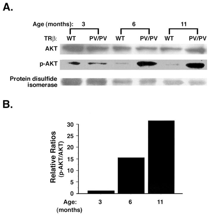
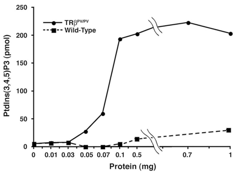
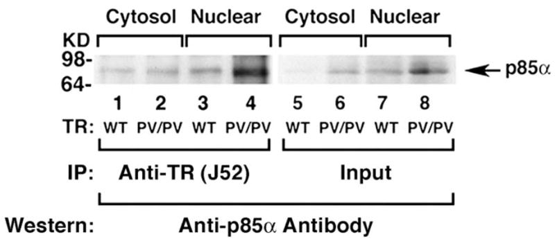
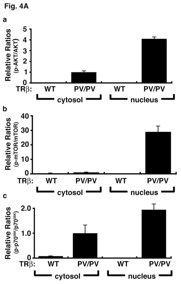
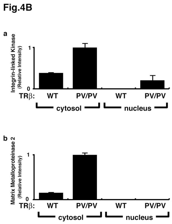
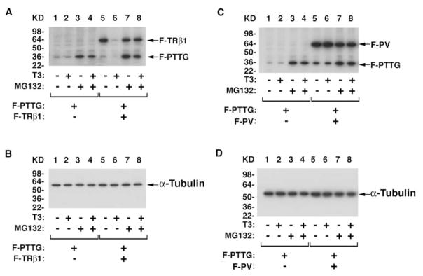
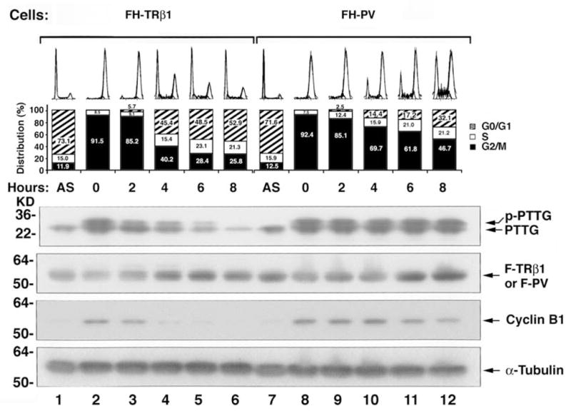
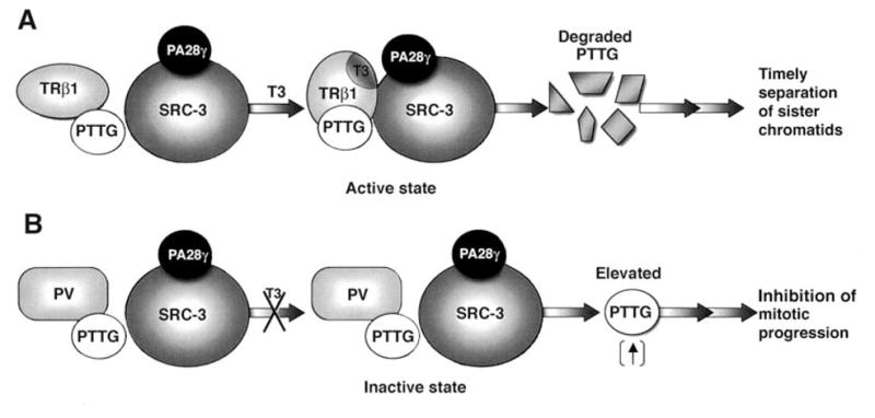
Study of molecular actions of thyroid hormone receptor beta (TRbeta) mutants in vivo has been facilitated by creation of a mouse model (TRbetaPV mouse) that harbors a knockin mutant of TRbeta (denoted PV). PV, which was identified in a patient with resistance to thyroid hormone, has lost T3 binding activity and transcription capacity. The striking phenotype of thyroid cancer exhibited by TRbeta(PV/PV) mice has allowed the elucidation of novel oncogenic activity of a TRbeta mutant (PV) [PAS1] beyond nucleus-initiated transcription. PV was found to physically interact with the regulatory p85alpha subunit of phosphatidylinositol 3-kinase (PI3K) in both the nuclear and cytoplasmic compartments. This protein-protein interaction activates the PI3K signaling by increasing phosphorylation of AKT, mammalian target of rapamycin (mTOR), and p70(S6K). PV, via interaction with p85alpha, also activates the PI3K-integrin-linked kinase-matrix metalloproteinase-2 signaling pathway in the extra-nuclear compartment. The PV-mediated PI3K activation results in increased cell proliferation, motility, migration, and metastasis. In addition to affecting these membrane-initiated signaling events, PV affects the stability of the pituitary tumor-transforming gene (PTTG) product. PTTG (also known as securin), a critical mitotic checkpoint protein, is physically associated with TRbeta or PV in vivo. Concomitant with T3-induced degradation of TRbeta, PTTG is degraded by the proteasome machinery, but no such degradation occurs when PTTG is associated with PV. The degradation of PTTG/TRbeta is activated by the direct interaction of the T3-bound TRbeta with the steroid receptor coactivator-3 (SRC-3) that recruits a proteasome activator (PA28gamma). PV that does not bind T3 cannot interact directly with SRC-3/PA28gamma to activate proteasome degradation, and the absence of degradation results in an aberrant accumulation of PTTG. The PV-induced failure of timely degradation of PTTG results in mitotic abnormalities. PV, via novel protein-protein interaction and transcription regulation, acts to antagonize the functions of wild-type TRs and contributes to the oncogenic functions of this mutation.


Figures

Fig. 1
Fig. 1
Activation of AKT by increased phosphorylation in the thyroids of TRβPV/PV mice in an age-dependent manner. (A). Thyroid extracts (50 μg) of TRβPV/PV mice and aged-matched wild-type mice were prepared and analyzed for total AKT, phosphorylated AKT (p-AKT), and protein disulfide isomerase (loading control) by Western blot analysis. (B). Densitometric analysis compared p-AKT to total AKT using a normalized ratio of TRβPV/PV mice (p-AKT/AKT)/wild-type mice (p-AKT/AKT) at each respective age [30].
Fig. 2
Fig. 2
Activation of PI3K activity in the thyroids of TRβPV/PV mice. Increasing concentrations as marked of total thyroid extracts from wild-type mice (solid squares) and TRβPV/PV mice (solid circles) were immunoprecipitated with anti-p85α antibody. The PI3K activity of precipitates from each concentration was measured for the conversion of PtdIns(3,4,5)P2 to PtdIns(3,4,5)P3 by ELISA. The data are expressed as the relative production of PtdIns(3,4,5)P3 by each sample [31].
Fig. 3
Fig. 3
p85α interacts more avidly with PV than with TRβ1 in the cytosolic and nuclear compartments. The thyroid extracts of 12 wild-type mice or three TRβPV/PV mice were pooled and separated into nuclear or cytosolic fractions. The purity of each fraction was monitored by the respective markers, poly ADP-ribose polymerase for the nuclear fraction and α-tubulin for the cytosolic fraction. An equal amount of the nuclear or cytosolic fraction (100 μg proteins) were each immunoprecipitated with 5 μg of monoclonal anti-TR/PV antibody J52 followed by Western blot analysis using anti-p85α antibody. Lanes are as marked. Lanes 5 – 8 show the corresponding input of the p85α protein.
Fig. 4
Fig. 4
Activation of the PI3K-AKT-mTOR-p70S6K and the integrin-linked kinase (ILK)-MMP2 pathways in the thyroids of TRβPV/PV mice. Pooled extracts from thyroids of 12 wild-type mice or three TRβPV/PV mice were separated into nuclear and cytosolic fractions. The purity of the fractions was confirmed by using respective markers, poly ADP-ribose polymerase for the nuclear fraction and α-tubulin for the cytosolic fraction. (A). Western blot analysis was carried out to determine cytosolic and nuclear abundance of the following proteins: total AKT, p-AKT(S473), total mTOR, p-mTOR, total p70S6K, and p-p70S6K. The band intensities were quantified and the ratios of p-AKT/total AKT (a), p-mTOR/total mTOR (b), p-p70S6K/total p70S6K (c) were expressed to indicate the activation of the respective effectors. (B). Western blot analysis of protein abundance of ILK (a) and MMP2 (b) in the cytosolic fraction of thyroid tumors of TRβPV/PV mice. The activation of this pathway occurs mainly in the cytosolic compartment.
Fig. 4
Fig. 4
Activation of the PI3K-AKT-mTOR-p70S6K and the integrin-linked kinase (ILK)-MMP2 pathways in the thyroids of TRβPV/PV mice. Pooled extracts from thyroids of 12 wild-type mice or three TRβPV/PV mice were separated into nuclear and cytosolic fractions. The purity of the fractions was confirmed by using respective markers, poly ADP-ribose polymerase for the nuclear fraction and α-tubulin for the cytosolic fraction. (A). Western blot analysis was carried out to determine cytosolic and nuclear abundance of the following proteins: total AKT, p-AKT(S473), total mTOR, p-mTOR, total p70S6K, and p-p70S6K. The band intensities were quantified and the ratios of p-AKT/total AKT (a), p-mTOR/total mTOR (b), p-p70S6K/total p70S6K (c) were expressed to indicate the activation of the respective effectors. (B). Western blot analysis of protein abundance of ILK (a) and MMP2 (b) in the cytosolic fraction of thyroid tumors of TRβPV/PV mice. The activation of this pathway occurs mainly in the cytosolic compartment.
Fig. 5
Fig. 5
T3-dependent proteasomal degradation of TRβ1 linked to the degradation of PTTG. Regulation of PTTG protein stability by the T3-bound TRβ1 via the proteasome-mediated pathway (A & B), but not by PV (C & D). [PAS10]CV1 cells were co-transfected with expression plasmids of F-PTTG, F-TRβ1 (A and B), F-PV (C and D) in the absence or presence (100 nM) of T3 and with or without MG132 as indicated. Antibodies used for Western blot analysis were: anti-Flag antibodies, anti-PTTG antibodies and for loading controls, anti-α-tubulin (B and D) [49].
Fig. 6
Fig. 6
Delayed mitotic exit of cells stably expressing PV (FH-PV cells) as compared with cells stably expressing TRβ1 (FH-TRβ1 cells). Aberrant accumulation of PTTG induced by PV impedes mitotic progression. FH-TRβ1 and FH-PV cells were synchronized in prometaphase/metaphase by thymidine/nocodazole block. Synchronized cells were collected at time 0 hour and then released by changing to growth media containing 20% fetal bovine serum. Cells harvested at various time points after release were subjected to Western blot and FACS analyses. An asynchronous population of FH-TRβ1 and FH-PV cells was also examined (AS). The protein abundance of PTTG, F-TRβ1, F-PV, cyclin B1, and loading control, α-tubulin, was determined by Western blot analysis [49].
Fig. 7
Fig. 7
A proposed molecular model for the liganded TRβ1-dependent SRC-3/PA28γ activated degradation of PTTG (A). The direct interaction of TRβ1 with SRC-3 induced by the binding of T3 results in the formation of liganded TRβ1/PTTG/SRC-3/PA28γ complexes that activate the degradation of PTTG. The degradation of PTTG leads to timely separation of sister chromatids. (B). PV also forms complexes with PTTG. But because PV does not bind T3, PV cannot directly bind to SRC-3/PA28γ to activate degradation. The absence of degradation leads to elevated PTTG and results in inhibition of mitotic progression [49].

References

    1. Weinberger C, Thompson CC, Ong ES, Lebo R, Gruol DJ, Evans RM. The c-erb-A gene encodes a thyroid hormone receptor. Nature. 1986;324:641–6. - PubMed
    1. Sap J, Munoz A, Damm K, Goldberg Y, Ghysdael J, Leutz A, Beug H, Vennstrom B. The c-erb-A protein is a high-affinity receptor for thyroid hormone. Nature. 1986;324:635–40. - PubMed
    1. Bassett JH, Harvey CB, Williams GR. Mechanisms of thyroid hormone receptor-specific nuclear and extra nuclear actions. Mol Cell Endocrinol. 2003;213:1–11. - PubMed
    1. Yen PM, Ando S, Feng X, Liu Y, Maruvada P, Xia X. Thyroid hormone action at the cellular, genomic and target gene levels. Mol Cell Endocrinol. 2006;246:121–7. - PubMed
    1. Cheng SY. Multiple mechanisms for regulation of the transcriptional activity of thyroid hormone receptors. Rev Endocr Metab Disord. 2000;1:9–18. - PubMed

Publication types

Substances