. 2007 Mar;44(9):2184-94.
doi: 10.1016/j.molimm.2006.11.016. Epub 2006 Dec 12.

Cellular uptake followed by class I MHC presentation of some exogenous peptides contributes to T cell stimulatory capacity

Affiliations

Cellular uptake followed by class I MHC presentation of some exogenous peptides contributes to T cell stimulatory capacity

Susan E Brophy et al. Mol Immunol. 2007 Mar.

Abstract

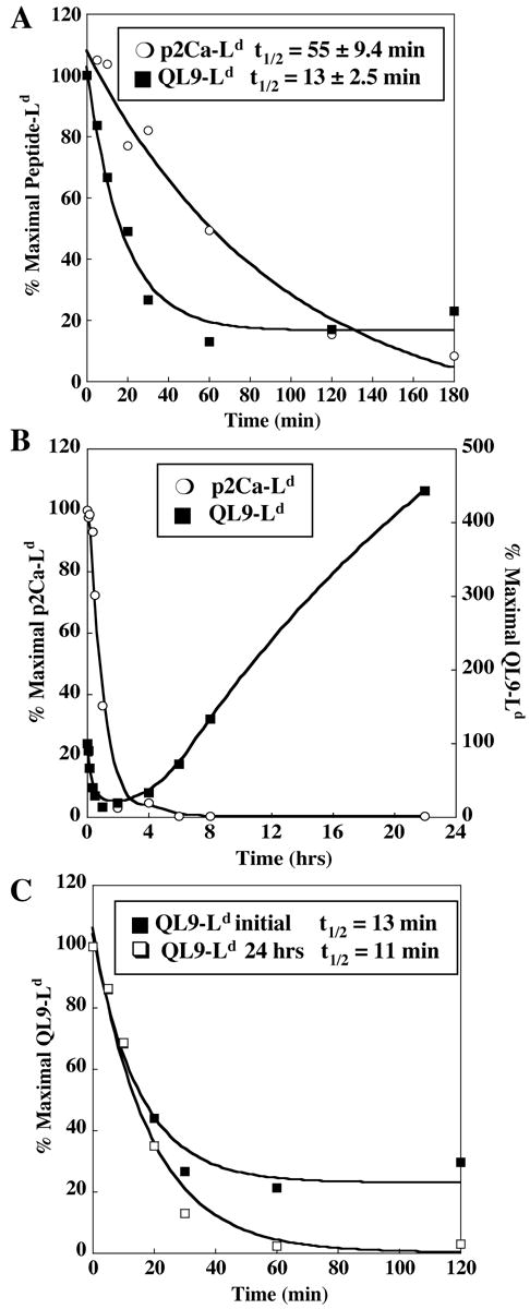
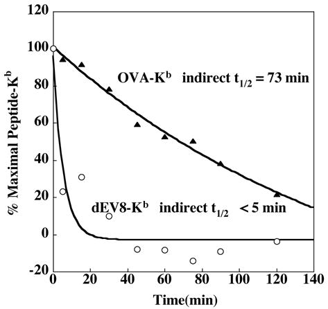
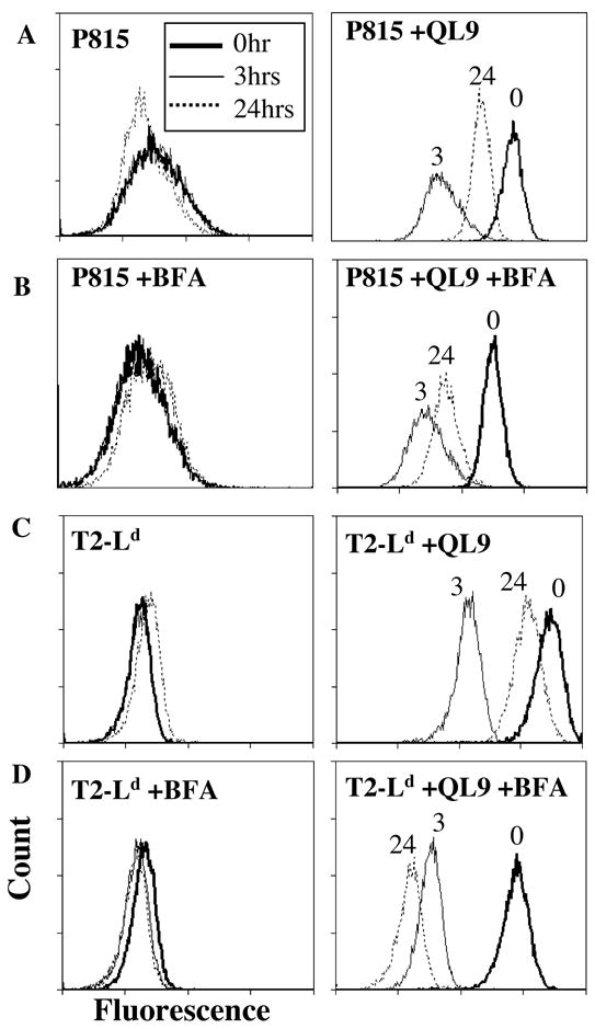
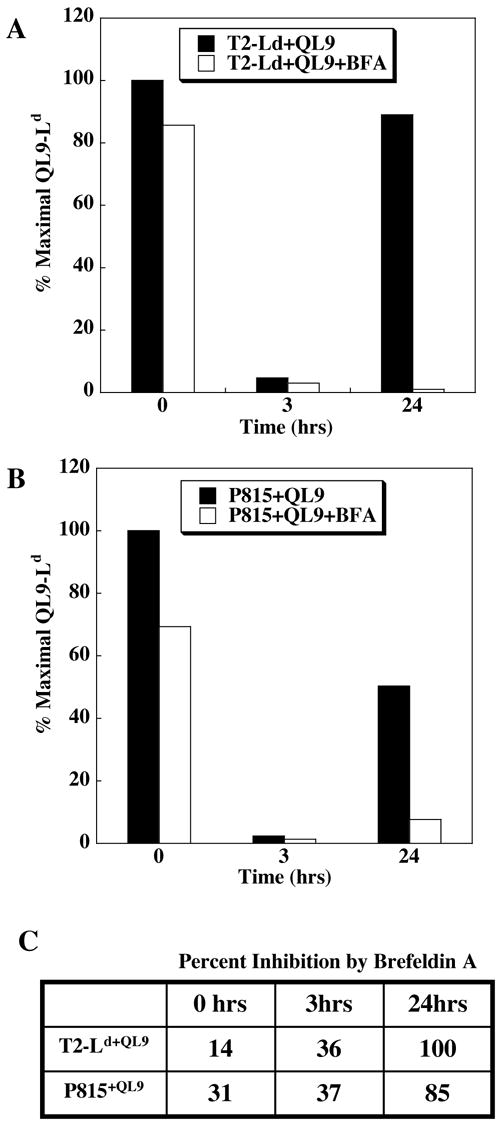
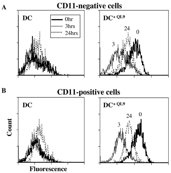
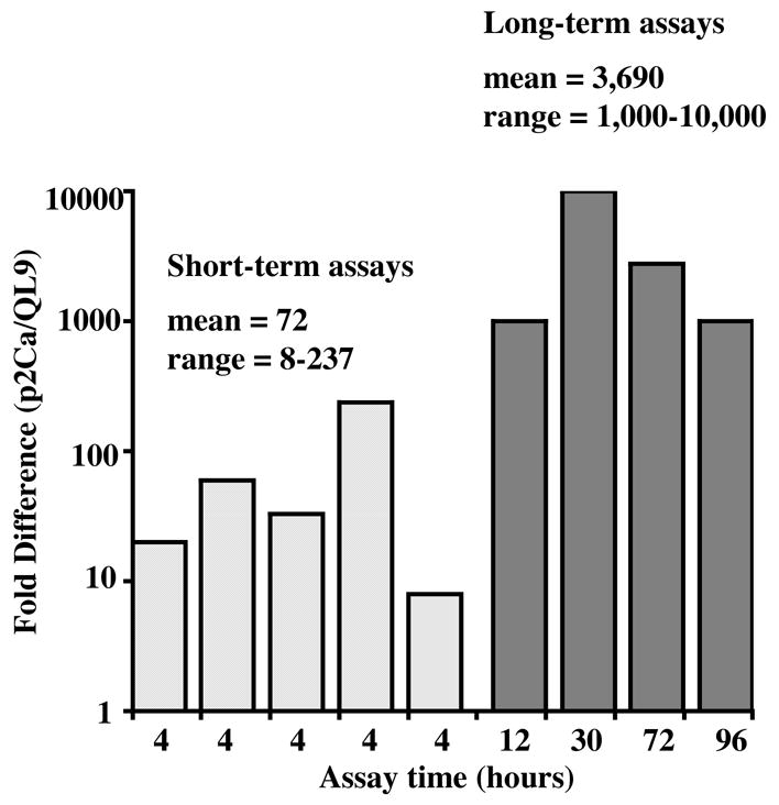
The T cell stimulatory activity of peptides is known to be associated with the cell surface stability and lifetime of the peptide-MHC (pepMHC) complex. In this report, soluble high-affinity T cell receptors (TCRs) that are specific for pepMHC complexes recognized by the mouse CD8+ clone 2C were used to monitor the cell surface lifetimes of synthetic agonist peptides. In the 2C system, L(d)-binding peptide p2Ca (LSPFPFDL) has up to 10,000-fold lower activity than peptide QL9 (QLSPFPFDL) even though the 2C TCR binds to p2Ca-L(d) and QL9-L(d) complexes with similar affinities. Unexpectedly, p2Ca-L(d) complexes were found to have a longer cell surface lifetime than QL9-L(d) complexes. However, the strong agonist activity of QL9 correlated with its ability to participate in efficient intracellular delivery followed by cell surface expression of the peptide, resulting in high and persistent surface levels of QL9-L(d). The ability of target cells to take up and present QL9 was observed with TAP-deficient cells and TAP-positive cells, including dendritic cells. The process was brefeldin A-sensitive, indicating a requirement for transport of the pepMHC through the ER and/or golgi. Thus, strong T cell stimulatory activity of some pepMHC complexes can be accomplished not only through long cell surface lifetimes of the ligand, but through a mechanism that leads to delayed presentation of the exogenous antigen after intracellular uptake.


Figures

FIGURE 1
FIGURE 1. Cell surface lifetimes of p2Ca-Ld and QL9-Ld
A. T2-Ld cells were incubated with p2Ca or QL9 peptide for 2 hours, washed, and suspended in media containing excess null peptide MCMV. Cells were incubated at 37°C and p2Ca-Ld and QL9-Ld levels were monitored at various time points by flow cytometry with m6α TCR. Standard deviation of cell surface lifetime was determined from four experiments. B. p2Ca-Ld and QL9-Ld levels were monitored over a 24 hour period, as described above. C. The initial QL9-Ld complexes (after the 2 hour incubation with peptide (see A above) and QL9-Ld complexes from the 24 hour time point in B were monitored in the presence of excess MCMV.
FIGURE 2
FIGURE 2. Cell surface lifetimes of dEV8-Kb and OVA-Kb
T2-Kb cells were incubated with dEV8, OVA, or without peptide for 2 hours. Cells were washed, resuspended in media containing excess SIYR peptide, and incubated at 37°C for various time periods. SIYR-Kb levels were detected by flow cytometry with m67α TCR. The amount of detectable SIYR-Kb complexes represented the loss of dEV8-Kb or OVA-Kb complexes. The percent maximal dEV8-Kb or OVA-Kb complex on the cell surface was calculated as described in Materials and Methods.
FIGURE 3
FIGURE 3. Cell surface levels of QL9-Ld on P815 and T2-Ld cells at various times, in the presence and absence of Brefeldin
A. P815 cells incubated without (A) or with (B) Brefeldin A (BFA) were incubated without or with QL9 peptide for 2 hours, washed and maintained at 37°C for 0 hours, 3 hours, or 24 hours. The level of QL9-Ld complexes was measured at these times using m6α TCR by flow cytometry. For BFA experiments, cells were treated 30 minutes prior to peptide incubation, after the 2 hour peptide pulse, and at 4 hours and 8 hours. T2-Ld cells were also incubated without (C) or with (D) Brefeldin A (BFA) and assayed as described for P815 cells.
FIGURE 4
FIGURE 4. Extent of inhibition of QL9-Ld surface levels by Brefeldin
A. Results from experiments described in Figure 3, were used to calculate the level of QL9-Ld relative to total Ld levels at different times for T2-Ld (A) or P815 (B). Bar graphs represent the percent QL9-Ld relative to the total amount of Ld on the cell surface at 0, 3, and 24 hours. The level of QL9-Ld and Ld on the cell surface was detected with m6α TCR and anti-Ld antibody 28.14.8, respectively. C. The percent inhibition of QL9-Ld complexes by BFA, relative to the absence of BFA, for T2-Ld and P815 cells pulsed with QL9 peptide.
FIGURE 5
FIGURE 5. Cell surface levels of QL9-Ld on bone marrow-derived dendritic cells at various times
Bone marrow cells were cultured in GM-CSF. On day 6, cells were washed, incubated with or without QL9 peptide for 2 hours, washed, and maintained at 37°C for 0 hours (bold line), 3 hours (solid line), and 24 hours (dashed line). QL9-Ld complexes were monitored with m6α TCR, gaiting on the CD11c-negative (A) and CD11c-positive (B) cell populations.
FIGURE 6
FIGURE 6. Relative activity of QL9 and p2Ca peptides in short and long-term assays
SD50 values from references listed in Table 2 were plotted as fold difference in SD50 values (p2Ca/QL9) versus assay time. Short-term assays (4 hours) are shown by the light grey bars and long-term assays (12–96 hours) are shown by the dark grey bars. Unpaired t-test (one-tailed) comparing the short-term and long-term values yielded a P value of 0.048.

Similar articles

Cited by

References

    1. Ackerman AL, Cresswell P. Cellular mechanisms governing cross-presentation of exogenous antigens. Nat Immunol. 2004;5:678–84. - PubMed
    1. Ackerman AL, Kyritsis C, Tampe R, Cresswell P. Access of soluble antigens to the endoplasmic reticulum can explain cross-presentation by dendritic cells. Nat Immunol. 2005;6:107–13. - PubMed
    1. Alexander J, Payne JA, Murray R, Frelinger JA, Cresswell P. Differential transport requirements of HLA and H-2 class I glycoproteins. Immunogenetics. 1989;29:380–399. - PubMed
    1. Andersen PS, Stryhn A, Hansen BE, Fugger L, Engberg J, Buus S. A recombinant antibody with the antigen-specific, major histocompatibility complex-restricted specificity of T cells. Proc Natl Acad Sci. 1996;93:1820–24. - PMC - PubMed
    1. Baker MB, Gagnon JS, Biddison EW, Wiley CD. Conversion of a T cell antagonist into an agonist by repairing a defect in the TCR/peptide/MHC interface: implications for TCR signaling. Immunity. 2000;13:475–84. - PubMed

Publication types

MeSH terms