. 2008 Mar;29(3):319-28.
doi: 10.1016/j.neurobiolaging.2006.10.027. Epub 2006 Dec 13.

Alzheimer's presenilin 1 causes chromosome missegregation and aneuploidy

Affiliations

Alzheimer's presenilin 1 causes chromosome missegregation and aneuploidy

Debrah I Boeras et al. Neurobiol Aging. 2008 Mar.

Abstract

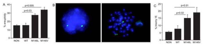
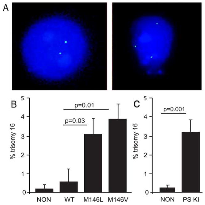
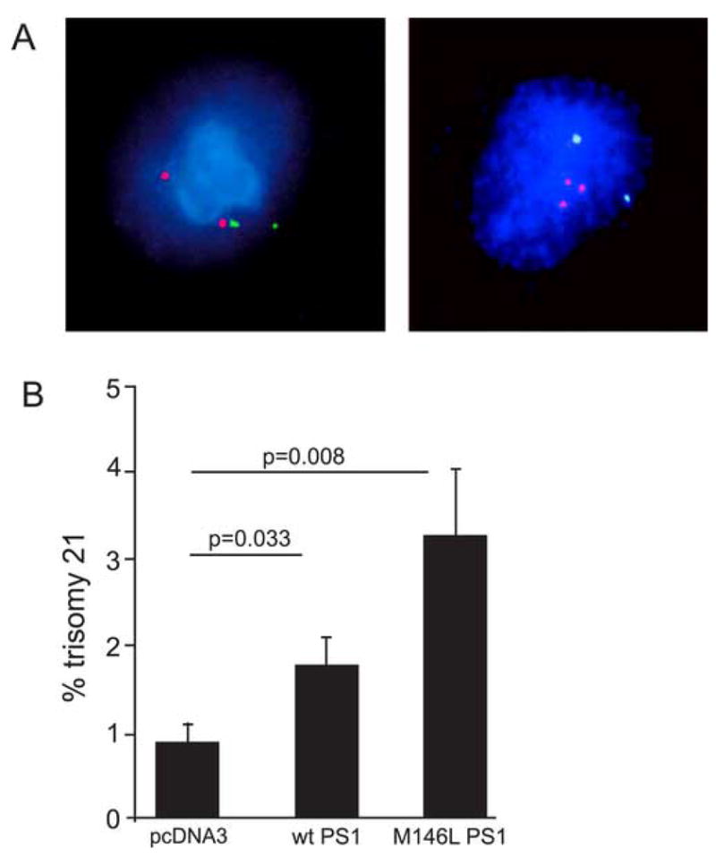
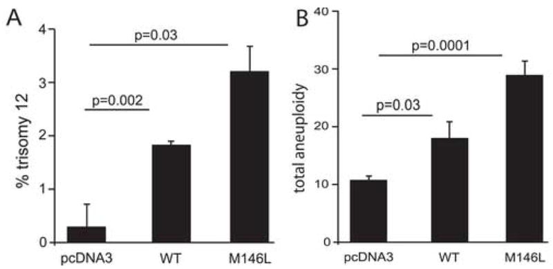
Mutations in the presenilin 1 gene cause most early onset familial Alzheimer's disease (FAD). Here, we report that a defect in the cell cycle - improper chromosome segregation - can be caused by abnormal presenilin function and therefore may contribute to AD pathogenesis. Specifically we find that either over-expression or FAD mutation in presenilin 1 (M146L and M146V) leads to chromosome missegregation and aneuploidy in vivo and in vitro: (1) Up to 20% of lymphocytes and neurons of FAD-PS-1 transgenic and knocking mice are aneuploid by metaphase chromosome analysis and in situ hybridization. (2) Transiently transfected human cells over-expressing normal or mutant PS-1 develop similar aneuploidy within 48 h, including trisomy 21. (3) Mitotic spindles in the PS-1 transfected cells contain abnormal microtubule arrays and lagging chromosomes. Several mechanisms by which chromosome missegregation induced by presenilin may contribute to Alzheimer's disease are discussed.


Conflict of interest statement

5.1 Disclosure Statement: No author has a conflict of interest. All Animal studies have been approved by the USF IACUC.

Figures

Figure 1
Figure 1
Aneuploidy induced in mouse spleen cells by a FAD mutant presenilin transgene. (A) Karyotype analysis of spleen cells of transgenic (WT: normal human PS-1; M146L: FAD mutant M146L PS-1; M146V: FAD mutant M146V PS-1) and non-transgenic mice (NON) revealed significantly higher levels of aneuploidy in the mutant PS-1 transgenic spleen cells. Mice were 15–17 months of age. (B) Fluorescence in situ hybridization (FISH) with a BAC probe (labeled with spectrum green) was used to score chromosome 16 in both interphase and metaphase spleen cells from mice transgenic for human PS-1 compared to nontransgenic mice. DAPI was used as a counter-stain. Diploid chromosome 16 is seen as two signals in the interphase cell on the left and seen as 4 signals, or 2 pairs of signals as in the metaphase cell on the right. (C) Quantitative in situ hybridization of spleen cells from transgenic and non-transgenic mice revealed significantly higher levels of trisomy 16 in the mutant PS-1 spleen cells. Mice were 14–19 months of age.
Figure 2
Figure 2
Aneuploidy induced in primary mouse brain neurons by a FAD PS-1 transgene. (A) FISH with a BAC probe (labeled with spectrum green) was used to score chromosome 16 in cultured neurons from PS-1 transgenic and non-transgenic mice. DAPI was used as a counter-stain. Chromosome 16 trisomy (right) reveals aneuploidy in neurons of PS-1 transgenic mice. Only 2 signals (disomy) were usually observed in neurons from non-transgenic mice (left). (B) Quantitative FISH of neurons from transgenic (WT: normal human PS-1; M146L: FAD mutant M146L PS-1; M146V: FAD mutant M146V PS-1) and non-transgenic mice (NON) revealed significantly higher levels of trisomy 16 only in the neurons from mice transgenic for FAD mutant PS-1. Mice were 14–19 months of age. (C) Quantitative FISH of cultured neurons from mutant (M146V) PS-1 Knock In (PS KI) and non-transgenic mice revealed significantly higher levels of trisomy 16 in the (M146V) PS-1 KI neurons. PS KI mice were 10–15 months of age and the non-transgenics were 17 months of age.
Figure 3
Figure 3
Increase in trisomy 21 aneuploidy in human cells induced by transfected normal and FAD mutant PS-1. (A) Examples of FISH with the LSI TEL/AML1 ES dual color probe used to detect both chromosome 12 (Spectrum Green) and 21 (Spectrum Orange) in PS-1 transfected hTERT cells. DAPI was used as a counter-stain. Chromosome 21 trisomy (right) reveals aneuploidy in a PS-1 transfected hTERT cell which is normal (disomic) for chromosome 12. A typical non-transfected hTERT cell (left) reveals normal (disomy) karyotype as evidenced by 2 red signals and 2 green signals. (B) Quantitative FISH of hTERT cells transfected with WT or mutant (M146L) PS-1 plasmids revealed significantly higher aneuploidy (trisomy 21) in both when compared to cells transfected with the empty vector.
Figure 4
Figure 4
PS-1-transfected human cells develop aneuploidy for many chromosomes. (A) Quantitative FISH on hTERT cells transiently transfected with WT or mutant (M146L) PS-1 revealed significant trisomy 12 aneuploidy when compared to empty vector control. (B) Karyotype analysis of hTERT cells transiently transfected with WT or mutant (M146L) PS-1 revealed significant aneuploidy when compared to cells transiently transfected with the empty vector. Up to 30% of cells became aneuploid 48 hours after transfection, and all size classes of chromosomes were affected.
Figure 5
Figure 5
Mitotic spindle abnormalities in PS-1 transfected cells. (A) hTERT cells were transfected for 48 hours with either the WT or M146L PS-1 plasmid or the empty vector and then stained with anti-α-tubulin monoclonal antibody and DAPI as a co-stain. Typical examples of cells in metaphase and anaphase are shown. Note the well-formed spindles, the correctly-positioned DNA and the clearly-defined microtubule organizing centers/centrosomes in the vector-transfected cells (a,b). In contrast, cultures transfected with PS-1-expressing plasmids developed numerous cells with abnormal mitotic spindles by 48 hours (c,d). For example, the first cell (c) appears to be in metaphase, however, there is no evidence that any centrosome or microtubule organizing center (MOC) has begun to develop, although the DNA is tightly arrayed along a metaphase plate. The next cell (d) appears to be in anaphase, but again the microtubule array and centrosomes are not well defined. Note also the apparent presence of lagging chromosomes left at the metaphase plate as the rest of the DNA has begun to move to opposite poles of the cell. (B) Quantification of the abnormal mitotic figures in cells transiently transfected with WT or mutant (M146L) PS-1 show more mitotic abnormalities (abnormal spindle appearance and lagging chromosomes) than cells transfected with the empty vector.

Similar articles

Cited by

References

    1. Arendt T, Rodel L, Gartner U, Holzer M. Expression of the cyclin-dependent kinase inhibitor p16 in alzheimer’s disease. Neuroreport. 1996;7(18):3047–3049. - PubMed
    1. Chevallier NL, Soriano S, Kang DE, Masliah E, Hu G, Koo EH. Perturbed neurogenesis in the adult hippocampus associated with presenilin-1 A246E mutation. Am J Pathol. 2005;167(1):151–159. - PMC - PubMed
    1. Chui DH, Tanahashi H, Ozawa K, Ikeda S, Checler F, Ueda O, Suzuki H, Araki W, Inoue H, Shirotani K, Takahashi K, Gallyas F, Tabira T. Transgenic mice with alzheimer presenilin 1 mutations show accelerated neurodegeneration without amyloid plaque formation. Nat Med. 1999;5(5):560–564. - PubMed
    1. Duesberg P. Are centrosomes or aneuploidy the key to cancer? Science. 1999;284(5423):2091–2092. - PubMed
    1. Duff K, Eckman C, Zehr C, Yu X, Prada CM, Perez-tur J, Hutton M, Buee L, Harigaya Y, Yager D, Morgan D, Gordon MN, Holcomb L, Refolo L, Zenk B, Hardy J, Younkin S. Increased amyloid-beta42(43) in brains of mice expressing mutant presenilin 1. Nature. 1996;383(6602):710–713. - PubMed

Publication types