. 2007;35(2):517-28.
doi: 10.1093/nar/gkl1080. Epub 2006 Dec 14.

Glucocorticoid-induced leucine zipper (GILZ)/NF-kappaB interaction: role of GILZ homo-dimerization and C-terminal domain

Affiliations

Glucocorticoid-induced leucine zipper (GILZ)/NF-kappaB interaction: role of GILZ homo-dimerization and C-terminal domain

Barbara Di Marco et al. Nucleic Acids Res. 2007.

Expression of concern in

Abstract

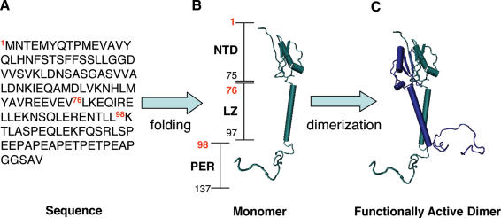
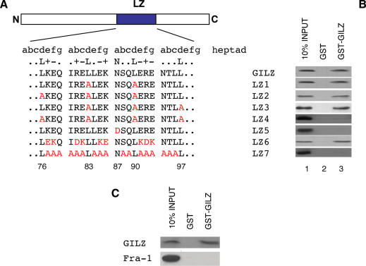
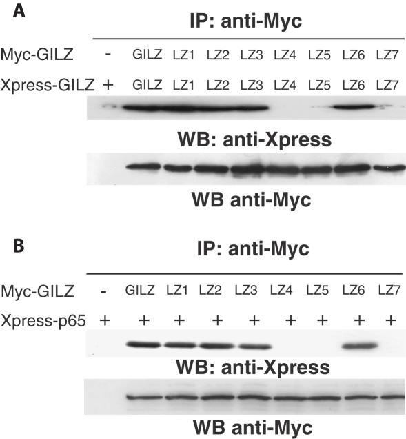
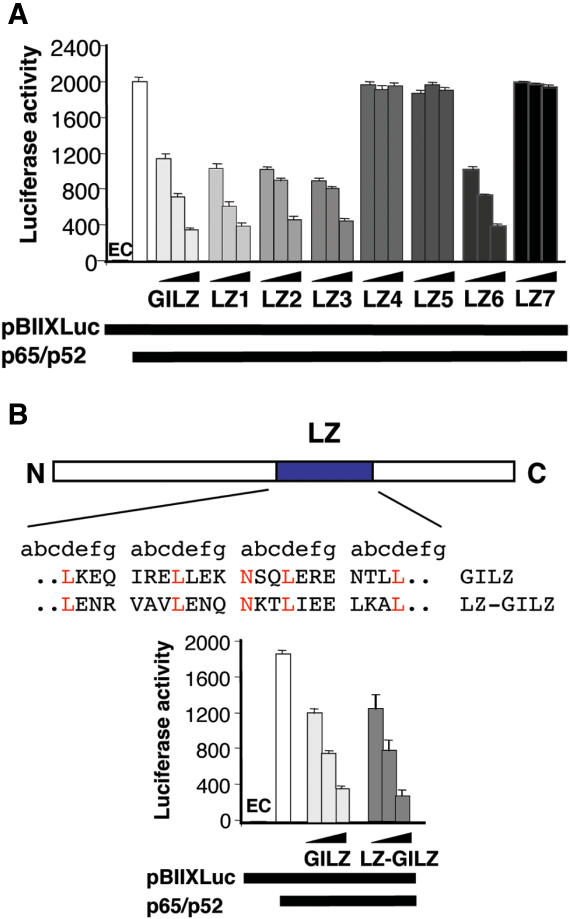
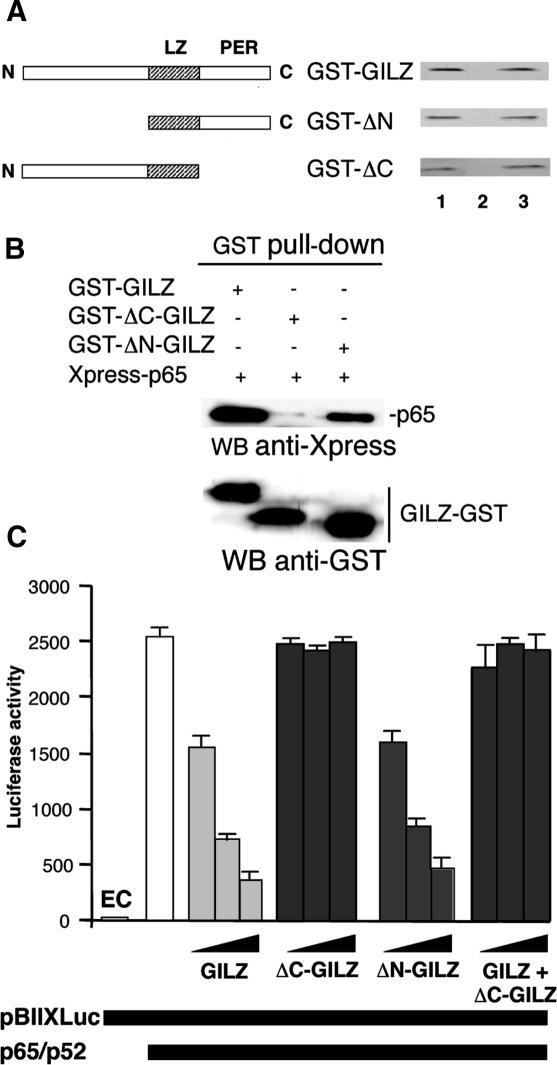
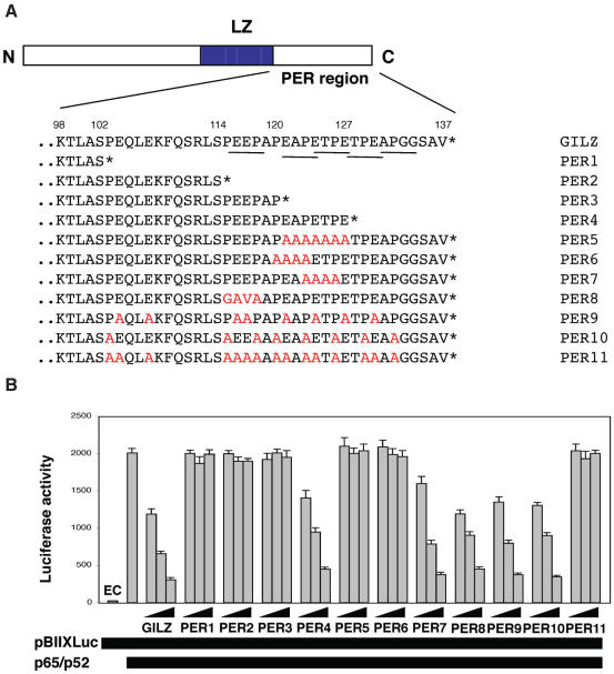
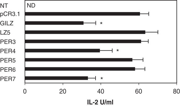
Glucocorticoid-induced leucine zipper (GILZ) is a 137 amino acid protein, rapidly induced by treatment with glucocorticoids (GC), characterized by a leucine zipper (LZ) domain (76-97 amino acids), an N-terminal domain (1-75 amino acids) and a C-terminal PER domain (98-137 amino acids) rich in proline and glutamic acid residues. We have previously shown that GILZ binds to and inhibits NF-kappaB activity. In the present study we used a number of mutants with the aim of defining the GILZ molecular domains responsible for GILZ/p65NF-kappaB interaction. Results, obtained by in vitro and in vivo co-immunoprecipitation (Co-IP) and by transcriptional activity experiments, indicate that GILZ homo-dimerization, through the LZ domain, as well as the C-terminal PER domain, particularly the 121-123 amino acids, are both necessary for GILZ interaction with NF-kappaB, inhibition of transcriptional activity and of IL-2 synthesis.


Figures

Figure 1
Figure 1
The sequence of 137 amino acid encoded by GILZ gene (A) is folded into a monomer (B), which is constituted by three domains: the N-terminal domain (NTD, 1–75 amino acids), the leucine zipper (LZ, 76–97 amino acids) and a C-terminal or PER domain (PER, 98–137 amino acids). Then, GILZ monomer is assembled into a functionally active dimer (C). The 3D model of the dimeric form of GILZ is obtained using the resolved structure of DIP as template [see Ref. (35)].
Figure 2
Figure 2
GILZ LZ domain is necessary for homo-dimerization in vitro. (A) Amino acid sequence of the LZ region of GILZ protein. LZ heptads are grouped (abcdefg) to help visualize LZ structure and mutations in this region are represented by red colored letters. Numbers in the bottom show amino acid position of GILZ protein relative to N-terminal region. (B) The 35S-labeled in vitro transcription/translation product of full-length wild-type GILZ was incubated with GST (column 2) or GST-GILZ or GST-GILZ mutants (column 3) immobilized on glutathione sepharose beads. The protein bound to the resin was eluted, resolved by SDS–PAGE, and visualized by autoradiography (right panel). 10% input indicates 0.1 vol of the 35S-labeled product used in the pull-down assay (column 1). The results are representative of one of three independent experiments. (C) The 35S-labeled in vitro transcription/translation product of full-length wild-type Fra-1 was incubated with GST (column 2) or GST-GILZ (column 3) immobilized on glutathione sepharose beads. The protein bound to the resin was eluted, resolved by SDS–PAGE, and visualized by autoradiography (right panel). 10% input indicates 0.1 volumes of the 35S-labeled product used in the pull-down assay (column 1). The results are representative of one of three independent experiments.
Figure 3
Figure 3
GILZ co-immunoprecipitates with NF-κB in Cos-1 cells and its homo-dimerization is necessary for GILZ–NF-κB interaction. (A) Lysates from Cos-1 cells transfected with 10 μg of vector containing Myc-tagged GILZ or its mutants (LZ1-7) and 10 μg of Xpress-tagged GILZ or its mutants (LZ1-7), were used for the IP analysis performed using anti-Myc antibody (Ab). Xpress-tagged GILZ or its mutants in the immunoprecipitates were detected by western blotting (WB) using anti-Xpress Ab. WB with anti-Myc Ab was performed to check Myc-tagged GILZ or its mutant's expression levels in transfected cells. (B) GILZ–p65NF-κB interaction was assessed by co-transfection in Cos-1 cells with 10 μg of vector containing Myc-tagged GILZ or its mutants (LZ1-7) and 10 μg of Xpress-tagged p65 protein. Xpress-tagged p65 in the immunoprecipitates were detected by WB using anti-Xpress Ab. WB with anti-Myc Ab was performed to check Myc-tagged GILZ or its mutants expression level in transfected cells.
Figure 4
Figure 4
(A) Homo-dimerization is important for GILZ inhibition of NF-κB transcriptional activity. Analysis of NF-κB transcriptional activity was performed in transiently co-transfected 293 cells. Different amounts of pCR3.1-GILZ (0.05, 0.5 and 5 μg) or its mutants, plus effector plasmids pCR3.1-p65 (1.5 μg) and pCR3.1-p52 (0.5 μg), were co-transfected with 15 μg of the reporter vector pBIIXLUC. Each transfection was performed in triplicate, and SD bars are shown. EC indicates endogenous control. (B) GILZ LZ domain was substituted with CREB LZ domain (LZ-GILZ, upper panel); NF-κB transcriptional activity was assessed by co-transfecting different amounts of pCR3.1-GILZ (0.05, 0.5 and 5 μg) or LZ-GILZ mutant, plus effector plasmids pCR3.1-p65 (1.5 μg) and pCR3.1-p52 (0.5 μg), together with 15 μg of the reporter vector pBIIXLUC. Each transfection was performed in triplicate, and SD bars are shown. EC indicates endogenous control.
Figure 5
Figure 5
Homo-dimerization is necessary but not sufficient for GILZ inhibition of NF-κB transcriptional activity. (A) Left panel: schematic representation of GILZ and its deleted mutants. Right panel: The 35S-labeled in vitro transcription/translation product of full-length wild-type GILZ was incubated with GST (column 2) or GST-GILZ or GST-GILZ mutants (column 3) immobilized on glutathione sepharose beads. The protein bound to the resin were eluted, resolved by SDS–PAGE, and visualized by autoradiography (right panel). 10% input used in the pull-down assay is shown in column 1. The results are representative of one of three independent experiments. (B) Lysates containing Xpress-p65 and GST–GILZ or GST–ΔC–GILZ or GST–ΔN–GILZ fusions proteins were pulled-down by glutathione Sepharose beads. Presence of fusions proteins was detected by WB with anti-GST Ab. Xpress-p65 precipitated was detected by WB with anti-Xpress Ab. (C) Analysis of NF-κB transcriptional activity was performed on transiently co-transfected 293 cells. Different amounts of pCR3.1-GILZ (0.05, 0.5 and 5 μg) or its deleted mutants, plus effector plasmids pCR3.1-p65 (1.5 μg) and pCR3.1-p52 (0.5 μ), were co-transfected with 15 μg of the reporter vector pBIIXLUC. In columns 11–13 (GILZ + ΔC-GILZ) equimolar pCR3.1-GILZ (0.025, 0.25 and 2.5 μg) and pCR3.1-ΔC-GILZ (0.025, 0.25 and 2.5 μg) plus effector plasmids pCR3.1-p65 (1.5 μg) and pCR3.1-p52 (0.5 μg), were co-transfected with 15 μg of the reporter vector pBIIXLUC. Each transfection was performed in triplicate, and SD bars are shown. EC indicates endogenous control.
Figure 6
Figure 6
Role of Pro/Glu rich (PER) region of GILZ in homo-dimerization and NF-κB transcriptional activity. (A) Amino acid sequence of the PER region of GILZ protein. The C-terminus is denoted with an asterisk. Point mutation is colored in red. PxxP domains are underscored. Numbers in the bottom show amino acid position of GILZ protein relative to the N-terminus. (B) Different amounts of pCR3.1-GILZ or GILZ mutants (0.05, 0.5 and 5 μg), plus effector plasmids pCR3.1-p65 (1.5 μg) and pCR3.1-p52 (0.5 μg), were co-transfected with 15 μg of the reporter vector pBIIXLUC. Each transfection was performed in triplicate, and SD bars are shown. EC indicates endogenous control.
Figure 7
Figure 7
(A) Backbone fluctuation of residues of PER region in the GILZ and PER9-11 mutants. Position 116, where the conformational bending of PER11 mutant starts, is marked with a red line. (B) Comparison of GILZ, PER9, PER10 and PER11 mutant conformations obtained at the end of the simulation. Alanine residues are shown in CPK style.
Figure 8
Figure 8
GILZ inhibits anti-CD3 IL-2 production. IL-2 production (U/ml) in untreated (NT) and anti-CD3 treated (1 μg/ml) for 18 h, empty vector transfected (pCR3.1) or GILZ or GILZ mutants (LZ5, PER3, PER4, PER5, PER6 and PER7) transfected 3DO cells as evaluated by ELISA assay. ND: not detected. Each transfection was performed in triplicate, and SD bars are shown. *P < 0.05.

References

    1. Iwata M., Hanaoka S., Sato K. Rescue of thymocytes and T cell hybridomas from glucocorticoid-induced apoptosis by stimulation via the T cell receptor/CD3 complex: a possible in vitro model for positive selection of the T cell repertoire. Eur. J. Immunol. 1991;21:643–648. - PubMed
    1. Kwak L.W., Longo D.L. Lymphomas. Cancer Chemother. Biol. Response Modif. 1996;16:376–440. - PubMed
    1. Raff M.C. Social controls on cell survival and cell death. Nature. 1992;356:397–400. - PubMed
    1. Frankfurt O., Rosen S.T. Mechanisms of glucocorticoid-induced apoptosis in hematologic malignancies: updates. Curr. Opin. Oncol. 2004;16:553–563. - PubMed
    1. Barnes P.J., Adcock I. Anti-inflammatory actions of steroids: molecular mechanisms. Trends Pharmacol. Sci. 1993;14:436–441. - PubMed

Publication types

MeSH terms