. 2007;35(2):477-85.
doi: 10.1093/nar/gkl1070. Epub 2006 Dec 14.

Selenium derivatization of nucleic acids for crystallography

Affiliations

Selenium derivatization of nucleic acids for crystallography

Jiansheng Jiang et al. Nucleic Acids Res. 2007.

Abstract

The high-resolution structure of the DNA (5'-GTGTACA-C-3') with the selenium derivatization at the 2'-position of T2 was determined via MAD and SAD phasing. The selenium-derivatized structure (1.28 A resolution) with the 2'-Se modification in the minor groove is isomorphorous to the native structure (2.0 A). To directly compare with the conventional bromine derivatization, we incorporated bromine into the 5-postion of T4, determined the bromine-derivatized DNA structure at 1.5 A resolution, and found that the local backbone torsion angles and solvent hydration patterns were altered in the structure with the Br incorporation in the major groove. Furthermore, while the native and Br-derivatized DNAs needed over a week to form reasonable-size crystals, we observed that the Se-derivatized DNAs grew crystals overnight with high-diffraction quality, suggesting that the Se derivatization facilitated the crystal formation. In addition, the Se-derivatized DNA sequences crystallized under a broader range of buffer conditions, and generally had a faster crystal growth rate. Our experimental results indicate that the selenium derivatization of DNAs may facilitate the determination of nucleic acid X-ray crystal structures in phasing and high-quality crystal growth. In addition, our results suggest that the Se derivatization can be an alternative to the conventional Br derivatization.


Figures
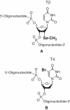
Figure 1
Figure 1
(A and B) Structures of the 2′-Se modification and 5-Br modification of DNA oligonucleotide.
Figure 2
Figure 2
Electron density maps and models of the derivatized DNA duplexes (Stereo view). Red and green balls represent Se and Br, respectively. Contours are at the 1.2σ level. (A) The structure of the Se-DNA (GdUSeGTACAC) at 1.28 Å resolution (1Z7I). (B) the structure of the Se-Br-DNA (GdUSeGdUBrACAC) at 1.5 Å resolution (2DLJ). (C) The structure of the Br-DNA (GTGdUBrACAC) at 1.8 Å resolution (2H05).
Figure 3
Figure 3
Electron density maps and models of the Se and Br modified nucleotides in the derivatized DNAs (Stereo view). Red and green balls represent Se and Br, respectively. Contours are at the 1.2σ level. (A) The structure of the 2′-Se-dU in d(GdUSeGTACAC) at 1.28 Å resolution (1Z7I). (B) The structure of the 5-Br-dU in d(GdUSeGdUBrACAC) at 1.50 Å resolution (2DLJ). (C) The structure of the 5-Br-dU in d(GTGdUBrACAC) at 1.80 Å resolution (2H05).
Figure 4
Figure 4
Superimposed comparison of the native and the derivatized DNA structures (red and green balls represent Se and Br, respectively). (A) The comparison of the Se-DNA (GdUSeGTACAC, 1Z7I in blue) and the native (GTGTACAC, 1DNS in pink). (B) The comparison of the Se-DNA dUse (1Z7I in blue) and the two native T2 (1DNS in pink and 1D78 in gray). (C) The comparison of the dUse of the Se-DNA (1Z7I in blue) and the reported DNA (GCGTAdUSeACGC, 1MA8 in yellow). (D) The comparison of the Br-DNA (GTGdUBrACAC, 2H05 in green) and the native (1DNS in pink). (E) The comparison of the Se-Br-DNA (GdUSeGdUBrACAC, 2DLJ in cyan) and the native (1DNS in pink).
Figure 5
Figure 5
The superimposed comparison of the native and Br-DNA local structures (green ball represents Br). The green and gray dash lines represent hydrogen and non-hydrogen bonds, respectively. (A) The comparison of the dUBr of the Br-DNA (GTGdUBrACAC, 2H05 in green, and water molecules in blue balls) and the T4 of the native DNA (GTGTACAC, 1DNS in pink). (B) The comparison of the dUBr of the Br-DNA (2H05 in green, and its water molecules in blue balls) and the T4 of the Se-DNA (GdUSeGTACAC, 1Z7I in blue, its water molecules in gray). (C) The comparison of the dG3 of the Br-DNA (in green) and the native (1DNS in pink). (D) The comparison of the T2 of the Br-DNA (in green) and the native (1DNS in pink).
Figure 6
Figure 6
The bromine hydration pattern and the local structures. Water molecules, Se, and Br are in blue, red and green balls, respectively. The green and gray dash lines represent hydrogen and non-hydrogen bonds, respectively. (A) The TGT backbone structure of the native DNA (GTGTACAC, 1D78). (B) The dUSeGT backbone structure of the Se-DNA (GdUSeGTACAC, 1Z7I). (C) The dUSeGdUBr backbone structure of the Se-Br-DNA structure (GdUSeGdUBrACAC, 2GPX). (D) The TGdUBr backbone structure of the Br-DNA (GTGdUBrACAC, 2H05).

Similar articles

Cited by

References

    1. MacRae I.J., Zhou K., Li F., Repic A., Brooks A.N., Cande W.Z., Adams P.D., Doudna J.A. Structural basis for double-stranded RNA processing by Dicer. Science. 2006;311:195–198. - PubMed
    1. Jacobs S.A., Podell E.R., Cech T.R. Crystal structure of the essential N-terminal domain of telomerase reverse transcriptase. Nature Struct. Mol. Biol. 2006;13:218–225. - PubMed
    1. Adams P.L., Stahley M.R., Kosek A.B., Wang J., Strobel S.A. Crystal structure of a self-splicing group I intron with both exons. Nature. 2004;430:45–50. - PubMed
    1. Parkinson G.N., Lee M.P.H., Neidle S. Crystal structure of parallel quadruplexes from human telomeric DNA. Nature. 2002;417:876–880. - PubMed
    1. Peersen O.B., Ruggles J.A., Schultz S.C. Dimeric structure of the Oxytricha nova telomere end-binding protein alpha-subunit bound to ssDNA. Nature Struct. Biol. 2002;9:182–187. - PubMed

Publication types