. 2006 Dec 26;103(52):19695-700.
doi: 10.1073/pnas.0609140103. Epub 2006 Dec 14.

Molecular identification of microsomal acyl-CoA:glycerol-3-phosphate acyltransferase, a key enzyme in de novo triacylglycerol synthesis

Affiliations

Molecular identification of microsomal acyl-CoA:glycerol-3-phosphate acyltransferase, a key enzyme in de novo triacylglycerol synthesis

Jingsong Cao et al. Proc Natl Acad Sci U S A. .

Abstract

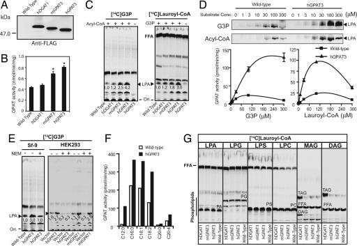
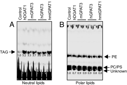
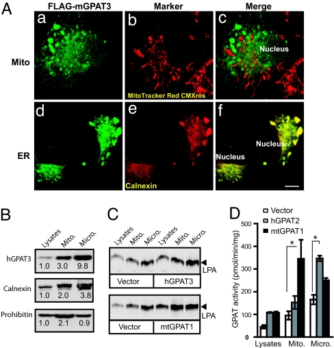
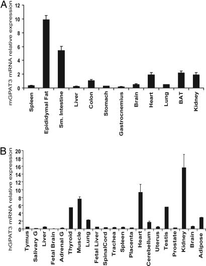
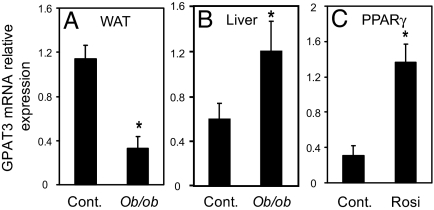
Acyl-CoA:glycerol-3-phosphate acyltransferase (GPAT) catalyzes the first step during de novo synthesis of triacylglycerol. It has been well recognized that mammals possess multiple enzymatically distinct proteins with GPAT activity. Although the mitochondrial-associated GPAT has been cloned and extensively characterized, the molecular identity of the endoplasmic reticulum (ER)-associated GPAT, which accounts for the majority of total GPAT activity in most tissues, has remained elusive. Here we report the identification of genes encoding human and mouse ER-associated GPAT (termed GPAT3). GPAT3 is a member of the acyltransferase family predominantly expressed in tissues characterized by active lipid metabolism, such as adipose tissue, small intestine, kidney, and heart. Ectopic expression of GPAT3 leads to a significant increase in N-ethylmaleimide-sensitive GPAT activity, whereas acyltransferase activity toward a variety of other lysophospholipids, as well as neutral lipid substrates, is not altered. Overexpression of GPAT3 in mammalian cells results in increased triacylglycerol, but not phospholipid, formation. GPAT3 is localized to the ER when overexpressed in COS-7 cells. GPAT3 mRNA is dramatically up-regulated during adipocyte differentiation, is reciprocally regulated in adipose tissue and liver of ob/ob mice, and is up-regulated in mice treated with a peroxisome proliferator-activated receptor gamma (PPARgamma) agonist. A substantial loss of GPAT activity in 3T3-L1 adipocytes was achieved by reducing GPAT3 mRNA levels through GPAT3-specific siRNA knockdown. These findings identify GPAT3 as a previously uncharacterized triacylglycerol biosynthetic enzyme. Similar to other lipogenic enzymes, GPAT3 may be useful as a target for the treatment of obesity.


Conflict of interest statement

Conflict of interest statement: H.F.L. owns publicly traded stock in Wyeth. The authors are employed by Wyeth and may also own publicly traded stock in Wyeth.

Figures

Fig. 1.
Fig. 1.
Sequence alignment of predicted acyltransferase domains of hGPAT3 (amino acids 209–332), human AGPAT1 (amino acids 83–211), and human mtGPAT1 (hGPAT1, amino acids 205–355). Underlined are four motifs (I–IV) characteristics of glycerophospholipid acyltransferases.
Fig. 2.
Fig. 2.
Enzymatic activity of GPAT3. Lysates from uninfected insect cells (wild type) or insect cells infected with virus expressing N-terminally FLAG-tagged human DGAT1 (hDGAT1) or native or N-terminally FLAG-tagged hGPAT3 and mGPAT3 were analyzed. In some experiments (E Right), lysates from mammalian HEK293 cells transfected with empty vector (vector) or hGPAT3- or mtGPAT1-containing vector were analyzed. (A) Western blot using anti-Flag antibody. (B) GPAT activity analyzed by butanol-extraction method (mean ± SD, n = 3). ∗, P < 0.05 compared with wild type or hDGAT1. (C) GPAT activity analyzed by TLC using either [14C]glycerol-3-phosphate (G3P, Left) or [14C]Lauroyl-CoA (Right) as radiolabeled substrates. The embedded numbers represent the relative levels of formed radiolabeled LPA (indicated by ▴). Ori, origin of migration; FFA, free fatty acid. The fast-migrating band next to LPA (C Left) may represent a G3P-dependent but acyl-CoA-independent product by endogenous enzyme(s), possibly phosphatidylglycerol phosphate or phosphatidylglycerol. Similar results were obtained in more than three independent experiments. (D) Substrate concentration dependence of GPAT activity. Assays were conducted by using Sf-9 lysates with the indicated concentrations of [14C]G3P or Lauroyl-CoA in the presence of 100 μM Lauroyl-CoA or [14C]G3P, respectively. Representative TLC images indicating the formation of LPA are shown on top; specific GPAT activities are shown below (mean ± SD, n = 3–4). (E) GPAT activity conferred by GPAT3, but not mtGPAT1, is sensitive to NEM. Enzymatic activity was analyzed in both Sf-9 cells (Left) and mammalian HEK293 cells (Right). (F) GPAT activity using different acyl-CoA species as substrates. Data represent the average of two independent experiments; variation between experiments was <15%. (G) Activity of GPAT3 toward different acyl acceptors. PA, phosphatidic acid; PG, phosphatidylglycerol; PS, phosphatidylserine; PC, phosphatidylcholine. Data are representative of two independent experiments with similar results.
Fig. 3.
Fig. 3.
Overexpression of GPAT3 in mammalian cells led to increased incorporation of oleic acid into TAG but not phospholipids. Metabolic labeling studies were performed in HEK293 cells overexpressing hGPAT3 and mGPAT3, human DGAT1, or human mtGPAT1 (hmtGPAT1) as described in Materials and Methods. (A) TLC analysis of neutral lipids. (B) TLC analysis of polar lipids. The number below each band represents relative level to that of the control, which was arbitrarily assigned as 1. Data are representative of two independent experiments with similar results. PE, phosphatidylethanolamine; PS, phosphatidylserine; PC, phosphatidylcholine.
Fig. 4.
Fig. 4.
GPAT3 is localized to ER but not mitochondria. (A) Immunofluorescent staining of COS-7 cells overexpressing Flag-tagged mGPAT3. GPAT3 is visualized by indirect immunofluorescence with anti-FLAG antibody (a and d), mitochondria and ER are visualized by staining with MitoTracker Red CMXRos (b) and antibody specific for Calnexin (e), respectively. c and f represent merged pictures of a with b and d with e, respectively. (Scale bar, 10 μm.) (B) Western blot analysis of subcellular fractions from HEK293 cells overexpressing FLAG-hGPAT3. The number below each band represents relative level to that of the lysates, which was arbitrarily assigned as 1. (C) TLC analysis of GPAT activity in subcellular fractions of HEK293 cells overexpressing FLAG-GPAT3 or mtGPAT1. (D) Quantitative analysis of GPAT activity (mean ± SD, n = 3–4). ∗, P < 0.05.
Fig. 5.
Fig. 5.
Tissue distribution of mouse (A) and human (B) GPAT3 mRNA detected by Q-PCR. Data are expressed as mean ± SD (n = 4).
Fig. 6.
Fig. 6.
GPAT3 mRNA expression and GPAT activity in 3T3-L1 adipocytes. (A) Induction of GPAT3 mRNA during 3T3-L1 differentiation detected by Q-PCR. (B and C) siRNA-mediated knockdown of GPAT3 in 3T3-L1 adipocytes. GPAT3 mRNA levels (B) and GPAT activity (C) were determined by Q-PCR analysis and TLC, respectively. Shown on top of C is a representative TLC datapoint showing the formation of LPA in the GPAT assay. Data are expressed as mean ± SD (n = 4). ∗, P < 0.05.
Fig. 7.
Fig. 7.
Regulation of GPAT3 mRNA in mice. (A and B) GPAT3 mRNA in white adipose tissue (WAT, A) or liver (B) of ob/ob mice compared with wild-type control mice. (C) Treatment of ob/ob mice with the PPARγ agonist rosiglitazone (Rosi) increased GPAT3 mRNA expression in WAT. Data are expressed as mean ± SEM. (n = 4). ∗, P < 0.05.

References

    1. Coleman RA, Lee DP. Prog Lipid Res. 2004;43:134–176. - PubMed
    1. Lehner R, Kuksis A. Prog Lipid Res. 1996;35:169–201. - PubMed
    1. Smith SJ, Cases S, Jensen DR, Chen HC, Sande E, Tow B, Sanan DA, Raber J, Eckel RH, Farese RV., Jr Nat Genet. 2000;25:87–90. - PubMed
    1. Stone SJ, Myers HM, Watkins SM, Brown BE, Feingold KR, Elias PM, Farese RV., Jr J Biol Chem. 2004;279:11767–11776. - PubMed
    1. Lewin TM, Schwerbrock NM, Lee DP, Coleman RA. J Biol Chem. 2004;279:13488–13495. - PubMed

MeSH terms