. 2007 Jan 10;26(1):242-52.
doi: 10.1038/sj.emboj.7601477. Epub 2006 Dec 14.

Unfolded protein response in a Drosophila model for retinal degeneration

Affiliations

Unfolded protein response in a Drosophila model for retinal degeneration

Hyung Don Ryoo et al. EMBO J. .

Abstract

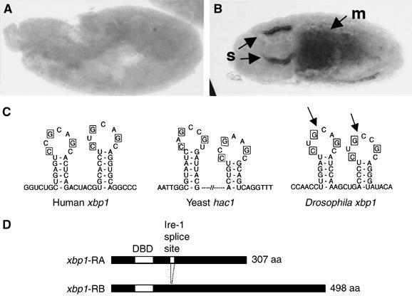
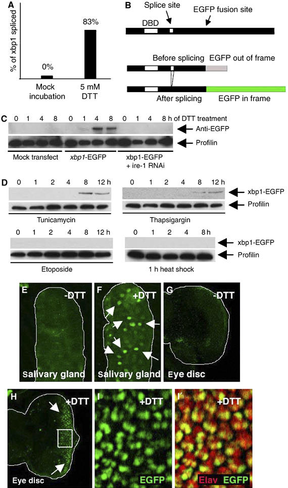
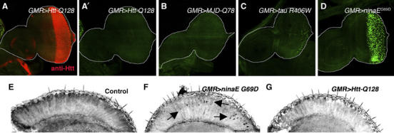
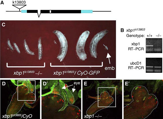
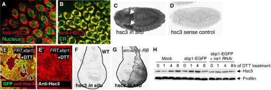
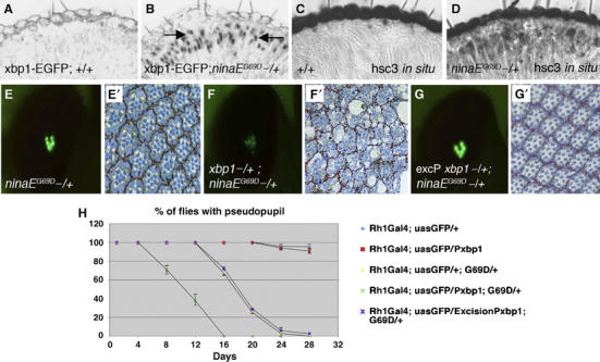
Stress in the endoplasmic reticulum (ER stress) and its cellular response, the unfolded protein response (UPR), are implicated in a wide variety of diseases, but its significance in many disorders remains to be validated in vivo. Here, we analyzed a branch of the UPR mediated by xbp1 in Drosophila to establish its role in neurodegenerative diseases. The Drosophila xbp1 mRNA undergoes ire-1-mediated unconventional splicing in response to ER stress, and this property was used to develop a specific UPR marker, xbp1-EGFP, in which EGFP is expressed in frame only after ER stress. xbp1-EGFP responds specifically to ER stress, but not to proteins that form cytoplasmic aggregates. The ire-1/xbp1 pathway regulates heat shock cognate protein 3 (hsc3), an ER chaperone. xbp1 splicing and hsc3 induction occur in the retina of ninaE(G69D)-/+, a Drosophila model for autosomal dominant retinitis pigmentosa (ADRP), and reduction of xbp1 gene dosage accelerates retinal degeneration of these animals. These results demonstrate the role of the UPR in the Drosophila ADRP model and open new opportunities for examining the UPR in other Drosophila disease models.


Figures

Figure 1
Figure 1
Structure and expression of Drosophila xbp1. (A, B) In situ hybridization against xbp1 mRNA in embryos. (A) A 6 h-old embryo shows little xbp1 mRNA. (B) A 12 h embryo with arrows pointing to high xbp1 mRNA in salivary glands (s) and the midgut (m). (C) A sequence comparison between the xbp1 genes of humans, Drosophila and the yeast hac1 shows a conserved stem–loop structure that is a target of Ire-1-mediated unconventional splicing. The bases required for Ire-1 recognition (in boxes) are conserved in the Drosophila xbp1 gene. The Ire-1 cleavage sites are marked with arrows. (D) The two predicted xbp1 isoforms. After Ire-1-mediated splicing, a frameshift in xbp1 translation occurs, converting a 307 aa protein into a 498 aa protein. The first white box indicates the DNA-binding domain (DBD). The second white box (smaller one) indicates the putative Ire-1 splice site.
Figure 2
Figure 2
ER stress activates xbp1 splicing. (A) The percentage of clones of xbp1-RB form sequenced from tissues mock incubated in Schneider's medium (left; n=17), and after exposure to 5 mM DTT (right; n=12). (B) The design of the xbp1-EGFP reporter. EGFP was inserted 3′ to the putative Ire-1 splice site, giving rise to a fusion protein that lacks the xbp1 C-terminal region (upper). EGFP is out of frame without Ire-1-mediated splicing (middle), but comes in frame after splicing (lower). (C) xbp1-EGFP marker is activated by Ire-1 in response to DTT treatment. The top panel shows anti-GFP Westerns whereas the lower panel shows anti-Profilin as a control. S2 cells with or without xbp1-EGFP transfection were incubated with 5 mM DTT for up to 8 h. xbp1-EGFP activation is seen at 1 h and peaks at 4 h. Pretreating these cells with ire-1 dsRNA abolishes xbp1-EGFP marker activation. (D) Specificity of xbp1-EGFP activation. S2 cells transfected with xbp1-EGFP were treated with chemicals or heat-shock stress. The upper panels show anti-GFP Westerns to detect xbp1-EGFP activation, whereas the lower panels show anti-Profilin blots as a loading control. The cells were incubated with tunicamycin (10 μg/ml), thapsigargin (2 μM) or etoposide (10 μM) in S2 medium for up to 12 h. For heat-shock stress, cells were heat shocked for 1 h (37°C) and allowed to recover for up to 8 h to resume protein synthesis. (E–I) Activation of the xbp1-EGFP marker in response to DTT (genotype: arm-Gal4/uas-xbp1-EGFP). EGFP is labeled in green. Activated xbp1-EGFP markers are indicated by arrows. (E, F) Third instar salivary glands that were mock incubated without DTT (E) or with 5 mM DTT for 3 h (F). (G, H) Eye imaginal discs that were mock incubated without DTT (G) or incubated with 5 mM DTT for 2 h (H). Spliced Xbp1-EGFP accumulates in the nucleus of these cells. (I) A higher magnification image of the inset in (H). Single channel of EGFP (I) and double labeling with anti-Elav (red) in (I′) show that xbp1-EGFP is more readily activated in photoreceptor cells.
Figure 3
Figure 3
xbp1-EGFP is activated in response to mutant Rh-1, but not to proteins that cause cytoplasmic aggregates. In all panels, proteins predicted to misfold were expressed in eye imaginal discs posterior to the morphogenetic furrow and in adult retinas by using the GMR-Gal4 driver. (A) Huntingtin-Q128 expression is visualized by staining with the anti-Htt antibody (red). (A′) The anti-GFP channel only (genotype: GMR-Gal4/uas-xbp1-EGFP; uas-Htt-Q128/+). (B) MJD-tr-Q78-expressing eye disc (genotype: GMR-Gal4/uas-xbp1-EGFP; uas-MJD-tr-Q78/+). (C) Tau R406W-expressing eye disc (genotype: GMR-Gal4/uas-xbp1-EGFP; uas-tau R406W/+). (D) Rh-1G69D-expressing eye disc shows strong xbp1-EGFP activation (genotype: GMR-Gal4/uas-xbp1-EGFP; uas-Rh-1G69D/+). (E–G) Adult eye horizontal sections. (E) A control adult head expressing ubcD1 (GMR-Gal4/uas-ubcD1; uas-xbp1-EGFP). (F) Rh-1G69D expression activates xbp1-EGFP in adult retinas (arrows). (G) By contrast, xbp1-EGFP activation was not detected in Htt-Q128-expressing retinas.
Figure 4
Figure 4
xbp1k13803−/− mutant phenotype. (A) The structure of xbp1 genomic locus. In the k13803 allele, a P-element is inserted 71 bp upstream of the putative transcription start site. The coding region is depicted in black boxes, the untranslated region in blue, one intron by a broken line and the unconventional ire-1 spliced intron in white. (B) RT–PCR reveals the loss of xbp1 transcripts in the xbp1k13803−/− larvae, compared to the wild-type controls (top). ubcD1, a ubiquitously expressed gene, is used as a loading control (bottom). (C) xbp1k13803 is a recessive lethal allele and shows larval growth retardation. Representative xbp1k13803−/− larvae are in the left and the xbp1/CyO (−/+) sibling control is in the right, at day 4 of development. Arrow points to an embryo (emb). (D, E) xbp1−/− larvae do not have imaginal discs. (D) Control larvae show eye imaginal discs on top of larval brains that are labeled with anti-Eya (red) and anti-Dronc (green; expressed ubiquitously in imaginal discs) (genotype: xbp1k13803/CyO-GFP). (D′) A magnified image of (D) in which eye discs are outlined and pointed with arrows. (E) xbp1−/− larvae do not have eye imaginal discs associated with larval brains (genotype: xbp1k13803/Df (2R)F36. (E′) A magnified image of the inset in (E).
Figure 5
Figure 5
Hsc3 is an ER chaperone regulated by the Ire-1/Xbp1 pathway. (A, B) Hsc3 localizes to the ER. (A) Co-labeling with Hsc3 (red) and the nuclear membrane marker, wheat germ agglutinin (green), shows perinuclear labeling. (B) Co-labeling with the ER-YFP marker (green) shows colocalization of Hsc3 to the ER compartment (genotype: sqh-ER-YFP). (C, D) hsc3 in situ hybridization in embryos. (C) hsc3 in situ signal in embryos. Arrows point to salivary glands. (D) hsc3 sense control does not label under identical conditions. (E) Hsc3 induction by DTT requires xbp1. An eye imaginal discs with eyeless-Flipase-induced clones of xbp1−/− are labeled by the absence of GFP (green) and outlined (white boundary) (genotype: eyFLP; FRT42D xbp1k13803/FRT42DubiGFP). The discs were treated with 5 mM DTT (in S2 cells medium) for 16 h. Upregulation of Hsc3 (red) occurs in the wild-type tissue but not in xbp1 mutant tissue. (F) In situ hybridization for hsc3 in wild-type wing imaginal discs. (G) hsc3 mRNA levels are enhanced in discs expressing xbp1-RB in the posterior compartment (right half of the disc and outlined; genotype: engrailed-Gal4/uas-xbp1-RB; tub-Gal80ts: induced at 29°C for 18 h). (H) Hsc3 induction by DTT treatment requires ire-1. The upper panel shows anti-Hsc3 Western blot, whereas the lower panel shows anti-Profilin as a loading control. While Hsc3 is induced by DTT in nontransfected or xbp1-EGFP-transfected cells, pretreatment of ire-1 dsRNA blocks Hsc3 induction.
Figure 6
Figure 6
ninaEG69D mutants activate the UPR, which suppresses the course of retinal degeneration. (A, B) Horizontal sections of 5- to 10–day-old adult eyes expressing xbp1-EGFP in a wild-type background (A) or in a ninaEG69D−/+ background (B). EGFP epitope appears in the nuclei of only the ninaEG69D−/+ outer photoreceptors (R1–R6, arrows). (C, D) The transcripts of hsc3 are at low basal level in wild-type (C), but are significantly enhanced in ninaEG69D−/+ retina (D). Genotypes are Rh-1-Gal4; uas-xbp1-EGFP/+ (in A, C) and Rh-1-Gal4; ninaEG69D/uas-xbp1-EGFP (in B, D). (E, F) Representative examples of the pseudopupil image visualized with Rh1Gal4; uasGFP, at experimental day 12, for ninaEG69D−/+ (E), xbp1k13803−/+; ninaEG69D−/+ (F) and a control in the background of a precisely excised chromosome of xbp1k13803, excision Pxbp1−/+; ninaEG69D−/+ (G). (E′–G′) Representative examples of the respective tangential sections of (E–G). (H) Quantification of the degeneration process by the pseudopupil assay. For each genotype, the percentage indicates the number of flies with pseudopupil/total number of flies. Each data point is an average of at least three independent assays. xbp1k13803 has a dominant effect in accelerating the retinal degeneration of ninaEG69D.

Similar articles

Cited by

References

    1. Armknecht S, Boutros M, Kiger A, Nybakken K, Mathey-Prevot B, Perrimon N (2005) High-throughput RNA interference screens in Drosophila tissue culture cells. Methods Enzymol 392: 55–73 - PubMed
    1. Back SH, Schroder M, Lee K, Zhang K, Kaufman RJ (2005) ER stress signaling by regulated splicing: IRE1/HAC1/XBP1. Methods 35: 395–416 - PubMed
    1. Bence NF, Sampat RM, Kopito RR (2001) Impairment of the ubiquitin–proteasome system by protein aggregation. Science 292: 1552–1555 - PubMed
    1. Bi M, Naczki C, Koritzinsky M, Fels D, Blais J, Hu N, Harding H, Novoa I, Varia M, Raleigh J, Scheuner D, Kaufman RJ, Bell J, Ron D, Wouters BG, Koumenis C (2005) ER stress-regulated translation increases tolerance to extreme hypoxia and promotes tumor growth. EMBO J 24: 3470–3481 - PMC - PubMed
    1. Brand AH, Perrimon N (1993) Targeted gene expression as a means of altering cell fates and generating dominant phenotypes. Development 118: 401–415 - PubMed

MeSH terms