. 2007 Jan 10;26(1):1-8.
doi: 10.1038/sj.emboj.7601466. Epub 2006 Dec 14.

Membrane potential governs lateral segregation of plasma membrane proteins and lipids in yeast

Affiliations

Membrane potential governs lateral segregation of plasma membrane proteins and lipids in yeast

Guido Grossmann et al. EMBO J. .

Abstract

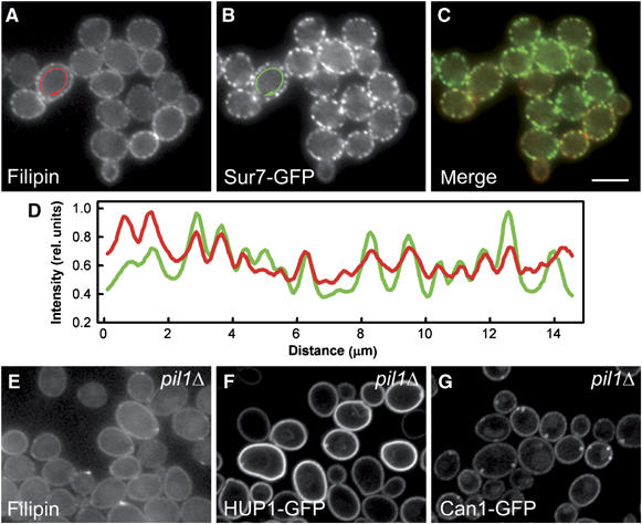
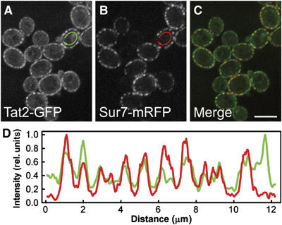
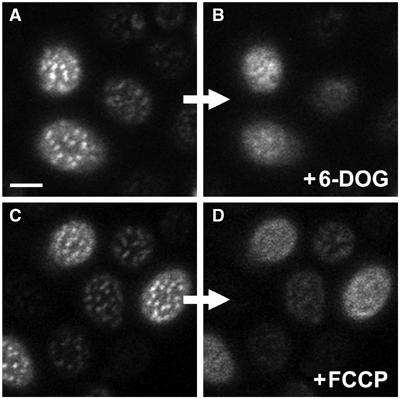
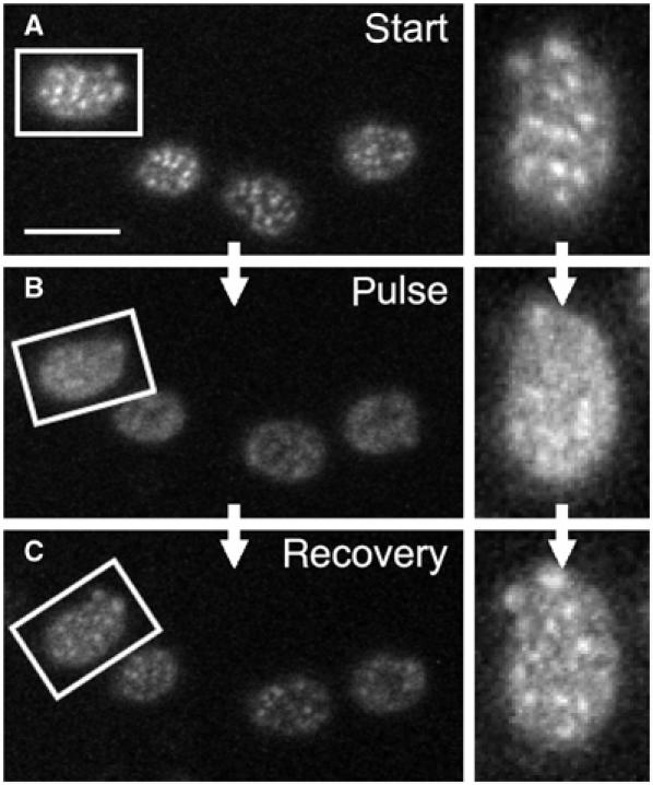
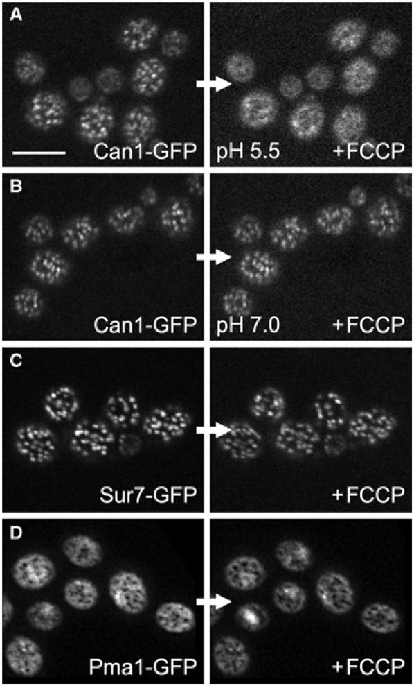
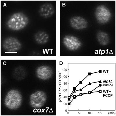
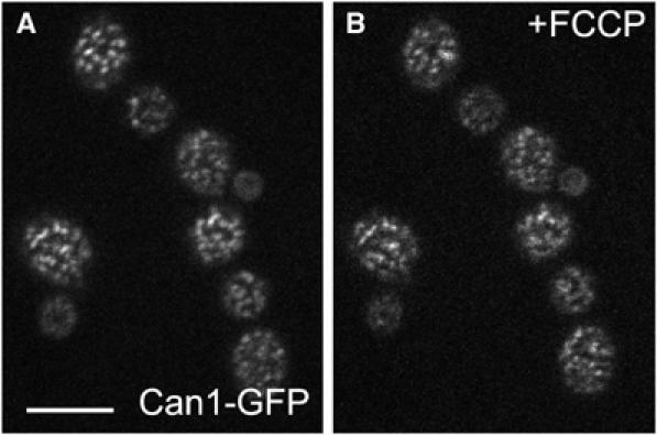
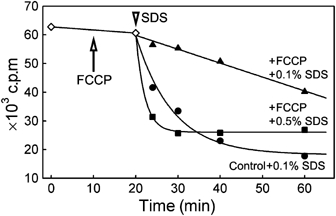
The plasma membrane potential is mainly considered as the driving force for ion and nutrient translocation. Using the yeast Saccharomyces cerevisiae as a model organism, we have discovered a novel role of the membrane potential in the organization of the plasma membrane. Within the yeast plasma membrane, two non-overlapping sub-compartments can be visualized. The first one, represented by a network-like structure, is occupied by the proton ATPase, Pma1, and the second one, forming 300-nm patches, houses a number of proton symporters (Can1, Fur4, Tat2 and HUP1) and Sur7, a component of the recently described eisosomes. Evidence is presented that sterols, the main lipid constituent of the plasma membrane, also accumulate within the patchy compartment. It is documented that this compartmentation is highly dependent on the energization of the membrane. Plasma membrane depolarization causes reversible dispersion of the H(+)-symporters, not however of the Sur7 protein. Mitochondrial mutants, affected in plasma membrane energization, show a significantly lower degree of membrane protein segregation. In accordance with these observations, depolarized membranes also considerably change their physical properties (detergent sensitivity).


Figures

Figure 1
Figure 1
Sites of sterol accumulation in plasma membrane colocalize with MCC. Simultaneous localization of filipin-stained sterols (A; red in C, D) and an MCC marker Sur7GFP (B; green in C, D) was performed in living GYS48 cells. Wide-field fluorescence micrographs (A–C) and the fluorescence intensity profiles along the cell surface (D; outside the arrows in A, B) are presented. The curves in (D) were smoothed using a mean filter to reduce the noise and normalized to the same maximum value. Distribution of filipin-stained sterols in pil1Δ mutant (GYS130; E) was compared with HUP1 (F) and Can1p (G) patterns in these cells (strains GYS131 and GYS132, respectively). Again, wide-field image is presented in (E), whereas confocal sections are shown in (F, G). Bar: 5 μm.
Figure 2
Figure 2
Tat2p is a part of MCC. Simultaneous localization of the tryptophan permease Tat2GFP (A; green in C) and the MCC marker Sur7mRFP (B; red in C) was performed in living GYS122 cells. Fluorescence intensity profiles along the cell surface (D; outside the arrows in A, B; green: Tat2GFP; red: Sur7mRFP) are presented. The curves in (D) were smoothed using a mean filter to reduce the noise and normalized to the same maximum value. Bar: 5 μm.
Figure 3
Figure 3
HUP1 patches are dissolved after plasma membrane depolarization. Changes in the distribution of HUP1GFP in the plasma membrane of living cells depolarized by 6-DOG uptake (strain GYS118; A, B) or FCCP treatment (strain GYS110; C, D; see Materials and methods for details) were observed. Surface confocal sections of the same cells before (A, C) and after the treatment (B, D) are presented. In both cases, the protein was released from MCC patches after plasma membrane depolarization. Only remnants of patches are visible in some treated cells. Bar: 2 μm. For corresponding time-lapse observations, see Supplementary Movies S1, S2 and S3.
Figure 4
Figure 4
Depolarization-induced changes in HUP1 distribution are reversible. For membrane depolarization, 1 min pulse of external electric field was applied on exponentially growing cells expressing HUP1GFP (strain GYS110) in order to depolarize the plasma membrane. The distribution of the protein before (A), immediately after (B) and 20 min after depolarization (C) is shown. Aligned surface sections of one cell are also presented (right). Despite the increased signal-to-noise ratio caused by phototobleaching of HUP1GFP fluorescence during the scanning, the restored pattern of MCC patches almost identical to that in (A) is clearly visible in (C). Bar: 5 μm.
Figure 5
Figure 5
Only some MCC proteins are affected by plasma membrane depolarization. The plasma membrane of living cells expressing Can1GFP (A, B), Sur7GFP (C) and Pma1GFP (D) (strains GYS113, GYS48 and KM12, respectively) was depolarized by FCCP. Surface optical sections of cells before (left) and after depolarization (right) are shown. Only Can1GFP pattern (A) was dissolved after the treatment. Neither Sur7GFP nor Pma1GFP plasma membrane distribution was affected (C, D). When the depolarization effect of FCCP was prevented using buffer of pH 7.0 during the treatment, no release of Can1p from MCC patches was observed (B). Bar: 5 μm.
Figure 6
Figure 6
The distribution of HUP1GFP is impaired in mutants deficient in mitochondrial respiration. Surface scans of WT cells (A), atp1Δ and cox7Δ mutants (B, C), all expressing HUP1GFP. For atp1Δ and cox7Δ, a considerably lowered membrane potential was detected by measurement of the uptake of 3H-TPP as compared to untreated and FCCP-treated WT cells (D). Bar: 2 μm.
Figure 7
Figure 7
Filipin stain prevents the release of Can1p from MCC. Cells expressing Can1GFP (strain GYS113) were stained by filipin and observed under agarose. These cells show the characteristic MCC pattern of Can1GFP fluorescence (A). To the same cells immobilized in agarose, 50 μM FCCP was added. The Can1-GFP pattern does not change (B) (compare with Can1-GFP cells depolarized by FCCP without filipin treatment; Figure 5A, right). Bar: 5 μm.
Figure 8
Figure 8
Leakage of AIB from the cells with depolarized plasma membrane. Cells accumulating radioactive AIB (strain BY4742) were treated with 0.1% SDS (arrowhead) in the absence (•) or presence of 20 μM FCCP (▴). The efflux of accumulated radioactivity was much slower in the presence of FCCP. Considerably higher detergent concentration (0.5% SDS; ▪) was necessary to induce an effect comparable to that observed in FCCP-untreated cells. Note that the addition of FCCP at t=10 min (arrow) had no effect on the membrane permeability for AIB (◊).

References

    1. Aggeler R, Capaldi RA (1990) Yeast cytochrome c oxidase subunit VII is essential for assembly of an active enzyme. Cloning, sequencing, and characterization of the nuclear-encoded gene. J Biol Chem 265: 16389–16393 - PubMed
    1. Anderson RGW, Jacobson K (2002) A role of lipid shells in targeting proteins to caveolae, rafts, and other lipid domains. Science 296: 1821–1825 - PubMed
    1. Bagnat M, Keranen S, Shevchenko A, Simons K (2000) Lipid rafts function in biosynthetic delivery of proteins to the cell surface in yeast. Proc Natl Acad Sci USA 97: 3254–3259 - PMC - PubMed
    1. Brachmann CB, Davies A, Cost GJ, Caputo E, Li J, Hieter P, Boeke JD (1998) Designer deletion strains derived from Saccharomyces cerevisiae S288C: a useful set of strains and plasmids for PCR-mediated gene disruption and other applications. Yeast 14: 115–132 - PubMed
    1. Brown DA, London E (1998) Functions of lipid rafts in biological membranes. Annu Rev Cell Dev Biol 14: 111–136 - PubMed

Publication types

MeSH terms