. 2007 Jan;8(1):84-90.
doi: 10.1038/sj.embor.7400855. Epub 2006 Dec 15.

Mdm2 targets the p53 transcription cofactor JMY for degradation

Affiliations

Mdm2 targets the p53 transcription cofactor JMY for degradation

Amanda S Coutts et al. EMBO Rep. 2007 Jan.

Abstract

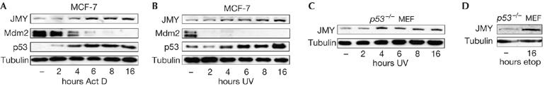
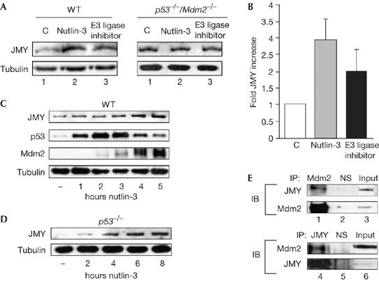
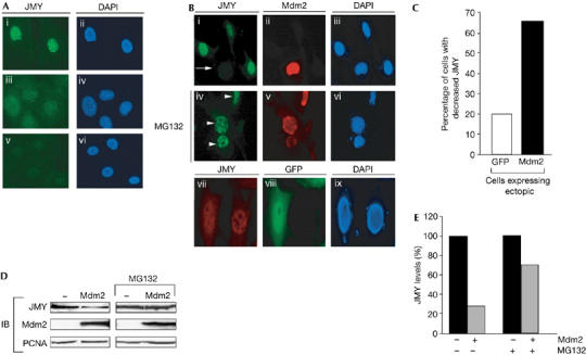
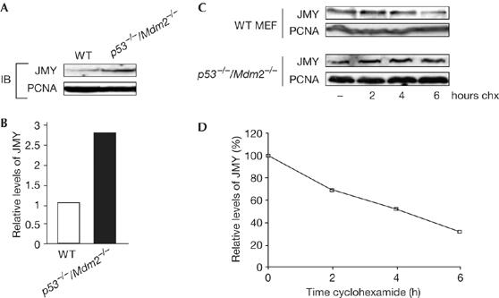
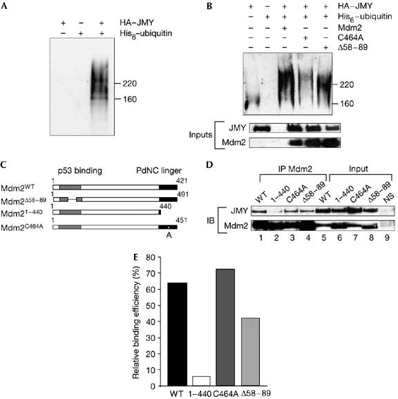
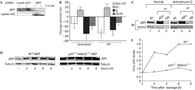
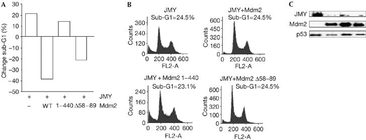
We define here a new mechanism through which Mdm2 (mouse double minute 2) regulates p53 activity, by targeting the p53 transcription cofactor JMY. DNA damage causes an increase in JMY protein, and, in a similar manner, small molecule inhibitors of Mdm2 activity induce JMY in unperturbed cells. At a mechanistic level, Mdm2 regulation of JMY requires the Mdm2 RING (really interesting new gene) finger, which promotes the ubiquitin-dependent degradation of JMY. However, regulation of JMY occurs independently of the p53-binding domain in Mdm2 and p53 activity. These results define a new functional relationship between the p53 cofactor JMY and Mdm2, and indicate that transcription cofactors that facilitate p53 activity are important targets for Mdm2 in suppressing the p53 response.


Figures

Figure 1
Figure 1
JMY is a DNA-damage responsive protein. (AD) Michigan Cancer Foundation-7 (MCF-7) or p53−/− mouse embryonic fibroblasts (MEFs) were treated with actinomycin D (Act D; 20 nM), ultraviolet (UV) light (50 J/m2), or etoposide (etop; 10 μM). Cell extracts were collected at the indicated time points and whole-cell extracts were run on 10% SDS–polyacrylamide gel electrophoresis. Endogenous JMY was detected using goat JMY antibody L-16.
Figure 2
Figure 2
JMY is regulated by mouse double minute 2. (A) Wild-type (WT) and p53−/−/Mdm2−/− mouse embryonic fibroblasts (MEFs) were treated as described with either nutlin-3 (10 μM) or Mdm2 E3 ligase inhibitor (10 μM) for 24 h and cell extracts were immunoblotted with JMY (upper panel) or tubulin (lower panel) antibodies. (B) The relative induction of JMY in WT MEFs compared with untreated cells (n=3, Mdm2 E3 ligase inhibitor; or n=4, nutlin-3; independent experiments). (C) WT MEFs were treated with 10 μM nutlin-3 for the times indicated. Endogenous JMY was detected using goat JMY antibody L-16. (D) p53−/− MEFs were treated with 10 μM nutlin-3 for the times indicated. Endogenous JMY was detected using goat JMY antibody L-16 and tubulin was used as a loading control. (E) Upper panels: endogenous Mdm2 was immunoprecipitated from whole-cell extracts prepared from monocyte chemoattractant factor-7 (MCF-7) cells by using mouse Mdm2 antibody SMP14 (lane 1). Mouse IgG was used as a nonspecific (NS) control (lane 2) and 2% of the extract was loaded in lane 3. Endogenous JMY was detected using goat JMY antibody L-16. Lower panels: endogenous JMY was immunoprecipitated from whole-cell extracts prepared from MCF-7 cells by using goat JMY antibody L-16 (lane 4). Goat IgG was used as an NS control (lane 5) and 2% of the extract was loaded in lane 6. IB, immunoblotting; IP, immunoprecipitation; Mdm2, mouse double minute 2.
Figure 3
Figure 3
Mouse double minute 2 reduces the level of nuclear JMY. (A) Endogenous JMY was detected in p53−/−/Mdm2−/− mouse embryonic fibroblasts (MEFs) using the JMY peptide antibody 1289 (i). Specific JMY staining was competed using the JMY peptide used to generate rabbit anti-serum (v). 4,6-Diamidino-2-phenylindole (DAPI) staining was used to visualize nuclei (ii, iv, vi). Nonspecific staining was detected using normal rabbit serum (iii). (B) p53−/−/Mdm2−/− MEFs were transfected with pCHDMIA encoding human Mdm2 and processed for immunofluorescence after 24 h. Endogenous JMY was detected using the rabbit anti-JMY antiserum 1289 (i, iv, vii). Exogenous human Mdm2 was detected using the mouse Mdm2 antibody SMP14 (ii, v). DAPI staining was performed to visualize nuclei (iii, vi, ix). The arrow indicates cells that express exogenous Mdm2 and show reduced levels of endogenous JMY. Where indicated (middle row; iv, v, vi), cells were treated with 10 μM MG132 for 8 h before fixation. Arrowheads indicate cells that express exogenous Mdm2 but do not show reduced JMY. As a control treatment, cells were transfected with pEGFP-C1 encoding green fluorescent protein (GFP; vii, viii, ix) and the level of endogenous JMY was monitored (vii). (C) pCHDMIA (expressing Mdm2) or pEGFP-C1 expression vector (500 ng) was transfected into p53−/−/Mdm2−/− MEFs grown on glass coverslips. Cells were stained with rabbit anti-JMY antiserum 1289 and Mdm2 antibody SMP14 or rabbit anti-JMY antiserum only for GFP-expressing cells. Fields of cells were compared for the level of JMY expression in the presence or absence of Mdm2 or GFP. The histogram represents the percentage of cells that had a decrease in JMY staining compared with surrounding cells. Approximately 50 cells were counted for each treatment. (D) p53−/−/Mdm2−/− MEFs were transfected with pCHDMIA encoding Mdm2 or an empty vector together with MG132 (10 μM) as indicated for 8 h before collecting. Endogenous JMY was detected using goat JMY antibody L-16 and Mdm2 was detected using mouse Mdm2 antibody SMP14. Proliferating-cell nuclear antigen (PCNA) was used as a loading control. (E) The percentage change in JMY levels compared with control treatments. IB, immunoblotting; Mdm2, mouse double minute 2.
Figure 4
Figure 4
JMY is regulated by a proteasome-dependent pathway. (A) Endogenous JMY was detected in wild-type (WT) and p53−/−/Mdm2−/− mouse embryonic fibroblasts (MEFs) using the JMY L-16 antibody; proliferating-cell nuclear antigen (PCNA) was used as a loading control. (B) The fold increase in JMY level in p53−/−/Mdm2−/− compared with WT MEFs. (C) WT or p53−/−/Mdm2−/− MEFs were treated with 50 μg/ml cyclohexamide (chx) for the times indicated, and cell extracts immunoblotted with the JMY antibody L-16. PCNA was used as a loading control. (D) The relative JMY levels in WT MEFs as a percentage of total JMY in p53−/−/Mdm2−/− MEFs. IB, immunoblotting.
Figure 5
Figure 5
JMY is ubiquitinated. (A) U2OS cells were transfected with 1 μg each of haemagglutinin (HA)–JMY, His6–ubiquitin or HA–JMY and His6–ubiquitin as indicated. Cells were treated with MG132 (10 μM) 6 h before collection and isolation of His-tagged proteins was performed using Ni-NTA agarose. Elutes were run on 7.5% SDS–polyacrylamide gel electrophoresis (SDS-PAGE) and JMY was detected using the goat JMY L-16 antibody. (B) U2OS cells were transfected with 1 μg each of HA–JMY, His6–ubiquitin and Mdm2 (mouse double minute 2) and derivates as denoted. Cells were treated with MG132 (10 μM) 4 h before collection. His-tagged proteins were isolated using Ni-NTA agarose and eluates were run on 7.5% SDS–PAGE. JMY derivatives were detected using the goat JMY L-16 antibody. The lower panel represent 20% of inputs used in His-tagged protein isolation. Whole-cell extracts were run on 10% SDS–PAGE and HA–JMY was detected using HA11 and Mdm2 was detected using SMP14 antibodies. (C) Representation of human Mdm2 and mutant derivatives. (D) U2OS cells were transfected with expression vectors encoding JMY (1 μg) and Mdm2 (2 μg), 1–440 (1 μg), C464A (1 μg) or Δ58–89 (2 μg) to a total of 3.4 μg DNA with vector control, as indicated. Cells were collected after 36 h followed by immunoprecipitation (IP) with the Mdm2 SMP14 antibody (lanes 1–4). JMY was detected with HA antibody HA11 and Mdm2 with SMP14. Because of the increased stability of 1–440 and C464A (Kubbutat et al, 1999), input Mdm2 protein levels were normalized before immunoprecipitation and 10% of each extract is shown (lanes 5–8). Lane 9 shows a control immunoprecipitation performed with a nonspecific (NS) mouse antibody and immunoblotted (IB) with either anti-HA or SMP14. (E) Quantification of the results, shown in (D), for the interaction between JMY and the Mdm2 mutant derivatives. The level of immunoprecipitated JMY was calculated as a relative percentage of the immunoprecipitated Mdm2 derivative, which was given an arbitrary value of 100% throughout to minimize any differences resulting from expression levels.
Figure 6
Figure 6
JMY regulation during the DNA damage response. (A) U2OS cells were treated with either JMY or lamin short interfering RNA (siRNA) as indicated. After 72 h, cells were collected followed by immunoblotting with goat JMY antibody L-16 or lamin antibody as indicated. (B) U2OS cells were treated as described with either JMY or lamin siRNA. Seven hours before the collection of cells, they were given either fresh medium or were treated with ultraviolet (UV) light (50 J/m2) and given fresh medium. Cells were analysed by flow cytometry (approximately 20,000 cells). The graph represents the change (expressed as %) in cell-cycle parameters relative to the control lamin siRNA-treated cells. Results represent mean±s.e.m. (n=4 independent experiments). Statistical analysis was performed using the Student's t-test: *P<0.05, **P<0.01, ***P<0.001. (C) U2OS cells were transfected with expression vectors encoding JMY (10 μg) and Mdm2 (10 μg). Cells were left untreated or were treated with actinomycin D (20 nM for 16 h). The cell extracts were immunoprecipitated (IP) with mouse haemagglutinin (HA) antibody (lanes 2, 5) or with mouse Gal4 antibody as a control (lanes 3, 6; NS). Immunoprecipitates were analysed for Mdm2 binding. JMY was detected with a rabbit HA antibody (Y-11) and Mdm2 with the rabbit H-221 antibody. Lanes 1 and 4 show the input (IN; 10% of cell extract). (D) Wild-type (WT) and p53−/−/Mdm2−/− mouse embryonic fibroblasts (MEFs) were treated with UV light (50 J/m2) for the times indicated. Cell extracts were collected and whole-cell extracts were run using 10% SDS–polyacrylamide gel electrophoresis. Endogenous JMY was detected using the goat JMY antibody L-16 and tubulin was used as a loading control. (E) The relative induction of JMY compared with untreated cells. Quantification was performed after normalizing JMY levels compared with loading controls. IB, immunoblotting.
Figure 7
Figure 7
Mouse double minute 2 regulates the functional properties of JMY. (A) U2OS cells were transfected with expression vectors encoding JMY together with expression vectors encoding mouse double minute 2 (Mdm2), Δ58–89 or 1–440, and collected after 48 h. The cell-cycle profile of approximately 10,000 cells was analysed by flow cytometry and the graph represents the relative change (expressed as a %) in sub-G1 cells relative to the control vector treatment. The graphs in (B) show representative fluorescence-activated cell sorting profiles with the total percentage of sub-G1 cells shown for each treatment. The panels in (C) show protein levels for each treatment. Ectopic JMY was detected using HA11, ectopic Mdm2 with SMP14 and endogenous p53 with DO-1. WT, wild type.

References

    1. Alarcon-Vargas D, Ronai Z (2002) p53–Mdm2—the affair that never ends. Carcinogenesis 23: 541–547 - PubMed
    1. Ardley HC, Robinson PA (2005) E3 ubiquitin ligases. Essays Biochem 41: 15–30 - PubMed
    1. Ashcroft M, Taya Y, Vousden KH (2000) Stress signals utilize multiple pathways to stabilize p53. Mol Cell Biol 20: 3224–3233 - PMC - PubMed
    1. Barak Y, Juven T, Haffner R, Oren M (1993) mdm2 expression is induced by wild type p53 activity. EMBO J 12: 461–468 - PMC - PubMed
    1. Boyd SD, Tsai KY, Jacks T (2000) An intact HDM2 RING-finger domain is required for nuclear exclusion of p53. Nat Cell Biol 2: 563–568 - PubMed

Publication types