. 2006 Dec 26;103(52):19836-41.
doi: 10.1073/pnas.0609628104. Epub 2006 Dec 15.

Filamin A (FLNA) is required for cell-cell contact in vascular development and cardiac morphogenesis

Affiliations

Filamin A (FLNA) is required for cell-cell contact in vascular development and cardiac morphogenesis

Yuanyi Feng et al. Proc Natl Acad Sci U S A. .

Abstract

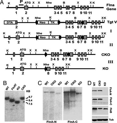
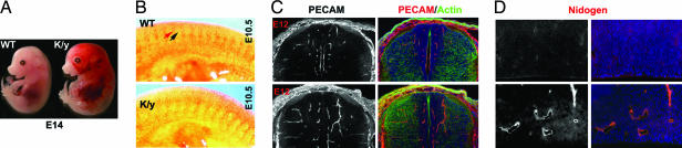
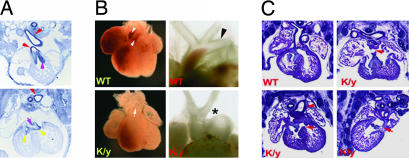
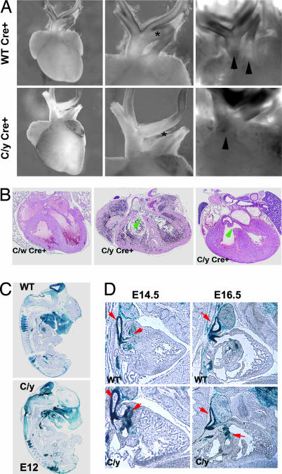
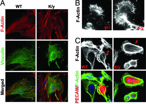
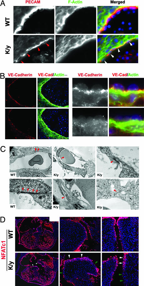
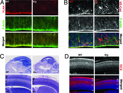
Mutations in the human Filamin A (FLNA) gene disrupt neuronal migration to the cerebral cortex and cause cardiovascular defects. Complete loss of Flna in mice results in embryonic lethality with severe cardiac structural defects involving ventricles, atria, and outflow tracts, as well as widespread aberrant vascular patterning. Despite these widespread developmental defects, migration and motility of many cell types does not appear to be affected. Instead, Flna-null embryos display abnormal epithelial and endothelial organization and aberrant adherens junctions in developing blood vessels, heart, brain, and other tissues. Essential roles for FLNA in intercellular junctions provide a mechanism for the diverse developmental defects seen in patients with FLNA mutations.


Conflict of interest statement

The authors declare no conflict of interest.

Figures

Fig. 1.
Fig. 1.
Targeting strategy for Flnac and null mutations. (A) A Neo-TK cassette flanked by loxP sites was inserted downstream of a 2.3-kb fragment containing exon 2 and intron 2 and upstream of an 8-kb fragment containing exons 3–11 of mouse Flna. A third loxP site was inserted into intron 7. The targeting vector was transfected into ES cells to generate Flna-targeted clones (Step I). Flna-recombinant ES cells were transfected with a Cre-recombinase-expressing plasmid, selected for ganciclovir resistance, and screened for deletion of the Neo-TK cassette alone (Step II) or with exons 3–7 of Flna (Step III). (B) A Southern blot analysis of Flna-targeted (FlnAT), FlnAK, and Flnac [conditional KO (CKO)] ES cells is shown. Genomic DNA was digested with KpnI. The Southern blot probe (P) is shown. The targeted Flna allele showed an 8.4-kb band instead of the 9.7-kb wild-type band. The 8.4-kb band reduced to 7.5 kb in the FlnaK allele and to 9.7 kb in the conditional allele. (C) Northern blot analysis of Flna-null and conditional-null ES cells. The blot was probed with N- and C-terminal portions of Flna cDNA. FlnaK cells showed a single Flna mRNA with deletion of exons 3–7. (D) An immunoblot of Flna mutant ES cells shows the deletion of Flna protein in the FlnaK allele and unaltered Flna protein in the Flnac allele (CKO). The blot was probed with antisera against peptides from mouse Flna N-terminal (anti-FLNA-N) and C-terminal (anti-FLNA-C) sequences. The blot was also probed with Flnb antiserum, which showed that Flnb expression is not increased in the absence of Flna. α-Tubulin (Tub) was used as loading control.
Fig. 2.
Fig. 2.
Vascular defects in Flna-null mice. (A) A dying E14 Flna-null mutant embryo and wild-type littermate. Note the widespread hemorrhage, dilated blood vessels, and edema in the Flna-null (K/y) mouse. (B) Immunostaining of E10.5 mouse embryo with PECAM antiserum shows disorganization of vasculature in the Flna-null mice (K/y). The red arrow indicates intersomitic vessels, and the black arrow indicates somites. Boundaries between somites and intersomitic vessels are missing in the Flna-null mutant. (C) Immunostaining of a spinal cord with PECAM antiserum (in red) at E12. Note the abundant and exuberant blood vessels in an FlnA mutant. Sections are costained with FITC-conjugated phalloidin (for F-actin in green) and Hoechst (in blue). (D) Immunostaining of E14 cortical ventricular epithelium with antisera to Nidogen (in red). Blood vessels in the Flna-null brain are dilated but show intact basement membranes.
Fig. 3.
Fig. 3.
Cardiac morphogenesis defects caused by Flna deficiency. (A) Flna expression in E12.5 mouse embryos is seen in the endocardial cushion (pink arrowheads); outflow tract (red arrowheads); and cardiac valves (yellow arrowheads). (B) Heart images of Flna-null (K/y) and wild-type littermates at E13.5 show the single outflow tract (PTA, indicated by the arrow) in the K/y mice. The K/y heart displayed PTA and interrupted aortic arch type B, with interruption between the left internal carotid artery and left subclavian artery (asterisk). Wild-type hearts show a normal aortic arch (arrowheads). (C) H&E-stained coronal sections of an Flna mutant heart (K/y) and of wild-type (Upper Left) littermates at E13.5. All Flna mutant embryos displayed incomplete septation of the ventricles (VSD, arrows) and outflow tracts (PTA, arrowhead). A single ventricle is seen (Upper Right), and combined atrial septal defect–VSD is seen (Lower Right).
Fig. 4.
Fig. 4.
Migration-independent neural crest defect in cardiac morphogenesis. (A) Heart images of Flna Wnt1-cre mutant males (C/y Cre+) and wild-type or heterozygous female litter mates. C/y Cre+ animals display PTA and interrupted aortic arch type B, with interruption between the left internal carotid artery and left subclavian artery (asterisk). (B) Pathological analysis (H&E stain) demonstrated that the outflow tract of male Wnt1-Cre Flnac mice (c/y Cre+) failed to septate, resulting in PTA (arrows). (C and D) Fate mapping of neural crest cells using ROZA26LacZ Cre reporter, in which the Flna mutant cells were visualized by expression of β-galactosidase. At E12.5, E14.5, and E16.5, Flna-deficient cells distribute normally in neural-crest-derived tissues, including the cardiac outflow tract and endocardial cushion (red arrows).
Fig. 5.
Fig. 5.
Normal F-actin structure, motility, and locomotion of Flna-null cells. (A) The immunofluorescence of mouse embryo fibroblasts from Flna-null embryos and littermates at E12.5. Cells were stained with a monoclonal antibody to Vinculin (in green) and costained with rhodamine-conjugated phalloidin (in red), and Hoechst (in blue). (B) The immunofluorescence of Flna-null neurons derived by differentiation of Flna-null ES cells with 0.5 μM retinoic acid or from cortical neural progenitor cells. Cells were stained with rhodamine-conjugated phalloidin to visualize F-actin in the neuronal growth cone. Cytoskeletal structures in Flna-null neurons are indistinguishable from wild-type neurons. (C) Flna-deficient endothelial cells identified by PECAM immunostaining (in red). F-actin intensity and structure visualized with FITC-conjugated phalloidin are similar in Flna-null and wild-type cells.
Fig. 6.
Fig. 6.
Defective AJs in Flna-deficient vascular endothelial cells. (A and B) Immunostaining with PECAM and VE-Cadherin antibodies showing that blood vessel endothelial cells are abnormally organized and that the intensity and distribution of VE-Cadherin are altered in the Flna-null mice at E12.5. Sections were costained with FITC-conjugated phalloidin (green) and Hoechst (blue). Arrows show disorganized and atrophic endothelial cells. (C) The transmission electron micrographs of capillary endothelial cells from Flna-null mice (K/y) and wild-type littermates. Arrowheads indicate AJs between neighboring cells. Note that the AJs in K/y endothelial cells are malformed with reduced electron density. (Magnification: ×1,000 or ×2,500, respectively.) (D) Defective endothelial cell organization in Flna-null hearts. Sections of developing hearts from Flna-null embryos and wild-type littermates at E12.5 show endothelial cells labeled with a monoclonal antibody to NFATc1 (in red). White arrowheads show endothelial cell clusters or multiple layers; green arrowheads indicate regions with absent or defective endothelial lining.
Fig. 7.
Fig. 7.
Brain development defects of Flna-null mice. (A) Immunostaining with a monoclonal antibody to Flna shows that Flna is concentrated at the apical surface of neuroepithelial cells in developing cerebral cortex. (B) Immunostaining with VE-Cadherin antibody (red) indicated its absence from the apical surface of the epithelial cells in cerebral cortex of Flna-null mice at E12.5. Sections also were costained with FITC-conjugated phalloidin (in green) and Hoechst (in blue). (C) Histological analysis of Flna-null mice (E14.5–E15) shows that the mutant brain was smaller, with reduced thickness in cortical plate and dilated blood vessels. (D) Immunostaining with Doublecortin (DCX) antiserum showed newly generated cortical neurons in the intermediate zone and cortical plate in Flna-null and wild-type brain, with no accumulation of neurons in cortical ventricular zone in Flna-null mutant. VZ, ventricular zone; IZ, intermediate zone; CP, cortical plate; MZ, marginal zone.

References

    1. Hartwig JH, Tyler J, Stossel TP. J Cell Biol. 1980;87:841–848. - PMC - PubMed
    1. Gorlin JB, Yamin R, Egan S, Stewart M, Stossel TP, Kwiatkowski DJ, Hartwig JH. J Cell Biol. 1990;111:1089–1105. - PMC - PubMed
    1. Feng Y, Walsh CA. Nat Cell Biol. 2004;6:1034–1038. - PubMed
    1. Stossel TP, Condeelis J, Cooley L, Hartwig JH, Noegel A, Schleicher M, Shapiro SS. Nat Rev Mol Cell Biol. 2001;2:138–145. - PubMed
    1. Eksioglu YZ, Scheffer IE, Cardenas P, Knoll J, DiMario F, Ramsby G, Berg M, Kamuro K, Berkovic SF, Duyk GM, et al. Neuron. 1996;16:77–87. - PubMed

Publication types

MeSH terms