. 2006 Dec 26;103(52):19713-8.
doi: 10.1073/pnas.0600943103. Epub 2006 Dec 15.

Topological dynamics of holins in programmed bacterial lysis

Affiliations

Topological dynamics of holins in programmed bacterial lysis

Taehyun Park et al. Proc Natl Acad Sci U S A. .

Abstract

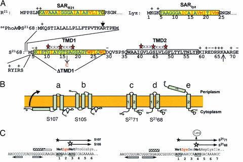
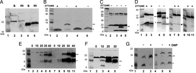
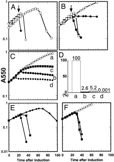
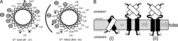
The fate of phage-infected bacteria is determined by the holin, a small membrane protein that triggers to disrupt the membrane at a programmed time, allowing a lysozyme to attack the cell wall. S(21)68, the holin of phage 21, has two transmembrane domains (TMDs) with a predicted N-in, C-in topology. Surprisingly, TMD1 of S(21)68 was found to be dispensable for function, to behave as a SAR ("signal-anchor-release") domain in exiting the membrane to the periplasm, and to engage in homotypic interactions in the soluble phase. The departure of TMD1 from the bilayer coincides with the lethal triggering of the holin and is accelerated by membrane depolarization. Basic residues added at the N terminus of S(21)68 prevent the escape of TMD1 to the periplasm and block hole formation by TMD2. Lysis thus depends on dynamic topology, in that removal of the inhibitory TMD1 from the bilayer frees TMD2 for programmed formation of lethal membrane lesions.


Conflict of interest statement

The authors declare no conflict of interest.

Figures

Fig. 1.
Fig. 1.
Properties of holins and holin genes. (A) Primary structure of S2168 and the N-terminal domains of Lyz, R21, and PhoA. The codon numbering for S2168 follows that of the full-length gene product, S2171 (see C for the dual-start structure at the beginning of S21). The SAR sequences of Lyz and R21 (7, 8) and TMD1 of S2168 (16) are shown in the yellow boxes, with the residues that are polar or neutral in terms of hydrophobicity in blue and the hydrophobic residues in red. TMD2 of S2168 is shown in an uncolored box. The red stars separated by three Xs above the TMDs of S21 indicate GxxxG-like motifs that may mediate interhelical interactions (19). The vertical arrow above the PhoA sequence indicates the normal signal sequence cleavage site (27). In ssPhoAΦS2168, the indicated sequence from PhoA is fused to the Met4 codon of S21. The position of the S16C missense change is indicated by a vertical arrow below the TMD1 sequence. The N terminus of S2168 was given two additional positive charges by inserting the sequence RYIRS between positions 4 and 5. To generate S2168ΔTMD1, the indicated sequence was deleted from S2168. The vertical arrow between residues 66 and 67 indicates the position where the sequence G2H6G2 was inserted in the allele used for purification. (B) Holin topologies. The topologies of the λ antiholin, S107 (a), and holin, S105 (b), are shown (6, 13, 23, 28). (c) and (d) show S2171 and S2168 with two TMDs, respectively. In (e), the lethal form of S21 is shown with its TMD1 in the periplasm. (C) Translational control region of the λ and 21 holin genes (11, 15, 16). Filled and empty stars show starts of the long (antiholin) and short (holin) gene products. Shine-Dalgarno sequences for the first and second translational starts of Sλ are indicated by striped and empty boxes, respectively. The single Shine–Dalgarno sequence of S21 is indicated by a striped box. The horizontal inverted pairs of arrows show the RNA stem loops controlling the dual starts. For S21, the vertical arrow shows the Met-4 → Leu mutation, which eliminates the production of the holin from the allele referred to as S2171. The Lys residues conferring antiholin character to the longer translational product in both the S and S21 genes are shown in red.
Fig. 2.
Fig. 2.
TMD1 exits the membrane. Aliquots from cultures expressing the indicated genes were collected by TCA precipitation and analyzed by SDS/PAGE under reducing or nonreducing conditions, as indicated. Except for A, where anti-Lyz antisera were used, separated proteins were detected by Western blotting using antisera raised against the C-terminal peptide of S21. Lane 1 in all panels, lanes 6 and 9 in D and lane 4 in G contain molecular mass standards. In BG, a star and a double star indicate the positions of the monomer and dimer forms of S2168S16C, respectively. (A) TMD1 of S2168 can substitute for the SAR domain of P1 Lyz. Lanes 2 and 3, S2168TMD1ΦLyzΔSAR; lanes 4 and 5, lyz; m, membrane fraction; s, soluble fraction. FtsI and Rλ were used as controls for the membrane and soluble fractions, respectively (see SI Fig. 6). (B) Dimerization of S2168S16C via its TMD1. Lanes 2 and 3, S2168S16C; lanes 4 and 5, RYIRSΦS2168S16C. Samples taken at 40 min after induction were prepared with or without the reducing agents DTT and β-mercaptoethanol, as indicated. (C) Disulfide formation reflects specific TMD1–TMD1 interactions. Lane 2, vector control; lane 3, S2168; lane 4, S2168S16C; lane 5, S2168G14C. (D) Protease sensitivity of S2168S16C dimers. In this panel, a large format Tris–Tricine gel system was used to allow resolution of the dimer- and monomer-related degradation products. Spheroplasts were prepared, induced for the expression of the indicated S2168 allele, and subsequently digested with proteinase K as described in Materials and Methods. Lanes 2–5, spheroplasts expressing S2168S16C and treated with protease for 0 (lanes 2 and 4) or 5 min (lanes 3 and 5); in lanes 2 and 3, the sample loading buffer contained reducing agents. Lanes 7–8, spheroplasts expressing RYIRSΦS2168S16C and treated with protease for 0 (lane 7) or 5 min (lane 8). Lanes 10–11, induced spheroplasts carrying the plasmid vector treated with protease for 0 (lane 10) or 5 min (lane 11). Symbols θθ and θ to the right of lane 5 indicate the position of the major degradation product of the dimer and monomer forms, respectively. A filled circle to the right of lane 8 indicates the position of the RYIRSΦS2168S16C monomer form. (E) Dimerization of S21 proteins increases as a function of time after induction. Lanes 2–6, S2168S16C; lanes 7–11, S2171S16C. Samples were taken at the times (minutes) indicated above the lanes and subjected to SDS/PAGE without reduction. (F) Direct export of TMD1 accelerates dimerization of the S21 holin. Lanes 2–5, ssphoAΦS2168S16C. Samples were taken at indicated times (minutes) and subjected to SDS/PAGE without reduction. (G) Collapse of the membrane potential accelerates dimerization of S21 proteins. Twenty minutes after induction, DNP was added to one of duplicate cultures expressing either S2168S16C or S2171S16C. Five minutes later, samples were taken from all four cultures and subjected to SDS/PAGE without reduction. Lanes 2 and 3, S2168S16C; lanes 5 and 6, S2171S16C.
Fig. 3.
Fig. 3.
The TMDs of S2168 have opposing functions. In each experiment, cultures of MG1655 lacIq1 tonA::Tn10 bearing a plasmid carrying the indicated S21 and R21 alleles under the control of the λ late promoter, pR′, and a compatible transactivating plasmid, pQ, carrying the λ late-activator gene Q under the control of a hybrid lac-ara promoter (see SI Table 1). The cultures were induced at time 0, and turbidity was followed as a function of time. Constructs used in A, B, E, and F contained the wild-type R21 gene, whereas those used in C carried, instead, R21E35Q, encoding an enzymatically inactive form of R21. (A) Expression of S2168 in combination with R21 results in abrupt host lysis. (●, ○), S2168 R21; ♦, S2168 amR21. DNP was added (arrow) to one culture (●) at 20 min after induction. (B) Expression of S2168ΔTMD1 in combination with R21 results in abrupt host lysis. (●, ○), vector control; (■, □), S2168ΔTMD1 R21; DNP was added (arrows) to two of the cultures (■, ○) at 25 min after induction. (C and D) Induction of different S2168 alleles in the absence of endolysin causes lethality. C shows growth curves after induction: (○, curve a), vector control; (■, curve b), S2168; (■, curve c), S2168ΔTMD1; (♦, curve d), ssphoAΦS2168. D shows cell survival at 60 min after induction, assessed as colony forming units (CFU) and expressed as a percentage of the control. (E) TMD1 of S2168 antagonizes the holin activity of its TMD2. (●), S2168 R21; (■), S2168S16C R21; (♦), RYIRSΦS2168S16C R21. (F) The SAR domain of P1 Lyz cannot inhibit hole formation by TMD2 of S2168. (○), SARlyzΦS2168ΔTMD1, uninduced; (●), SARlyzΦS2168ΔTMD1, induced; (■), RYIRS-SARlyzΦS2168ΔTMD1, induced.
Fig. 4.
Fig. 4.
Potential homotypic and heterotypic TMD interactions in the function of S21. (A) Helical projections of TMDs of S21. Stars indicate GxxxGxxxG-like motifs of each TMD (G10, G14, and G18 in TMD1; G40, S44, and G48 in TMD2). Hydrophilic or neutral residues in each TMD are circled and shaded. (Left) the SAR domain (TMD1) of S21 (residues 7–27), with the position of the S16C mutation highlighted by an asterisk. (Right) TMD2, the hole-forming domain of S21 (residues 36–60), with a potential hydrophilic surface that may line the lethal membrane lesion indicated by the arc. (B) Pathway to hole formation involves inhibited (i) and active (ii) topological isomers of S21. See Discussion for details.

References

    1. Fuhrman JA. Nature. 1999;399:541–548. - PubMed
    1. Hendrix RW, Smith MC, Burns RN, Ford ME, Hatfull GF. Proc Natl Acad Sci USA. 1999;96:2192–2197. - PMC - PubMed
    1. Young R, Wang IN, Roof WD. Trends Microbiol. 2000;8:120–128. - PubMed
    1. Young R. Microbiol Rev. 1992;56:430–481. - PMC - PubMed
    1. Gründling A, Manson MD, Young R. Proc Natl Acad Sci USA. 2001;98:9348–9352. - PMC - PubMed

Publication types

LinkOut - more resources