. 2006 Dec 26;103(52):19783-7.
doi: 10.1073/pnas.0609582104. Epub 2006 Dec 15.

Rae1 interaction with NuMA is required for bipolar spindle formation

Affiliations

Rae1 interaction with NuMA is required for bipolar spindle formation

Richard W Wong et al. Proc Natl Acad Sci U S A. .

Abstract

In eukaryotic cells, the faithful segregation of daughter chromosomes during cell division depends on formation of a microtubule (MT)-based bipolar spindle apparatus. The Nuclear Mitotic Apparatus protein (NuMA) is recruited from interphase nuclei to spindle MTs during mitosis. The carboxy terminal domain of NuMA binds MTs, allowing a NuMA dimer to function as a "divalent" crosslinker that bundles MTs. The messenger RNA export factor, Rae1, also binds to MTs. Lowering Rae1 or increasing NuMA levels in cells results in spindle abnormalities. We have identified a mitotic-specific interaction between Rae1 and NuMA and have explored the relationship between Rae1 and NuMA in spindle formation. We have mapped a specific binding site for Rae1 on NuMA that would convert a NuMA dimer to a "tetravalent" crosslinker of MTs. In mitosis, reducing Rae1 or increasing NuMA concentration would be expected to alter the valency of NuMA toward MTs; the "density" of NuMA-MT crosslinks in these conditions would be diminished, even though a threshold number of crosslinks sufficient to stabilize aberrant multipolar spindles may form. Consistent with this interpretation, we found that coupling NuMA overexpression to Rae1 overexpression or coupling Rae1 depletion to NuMA depletion prevented the formation of aberrant spindles. Likewise, we found that overexpression of the specific Rae1-binding domain of NuMA in HeLa cells led to aberrant spindle formation. These data point to the Rae1-NuMA interaction as a critical element for normal spindle formation in mitosis.


Conflict of interest statement

The authors declare no conflict of interest.

Figures

Fig. 1.
Fig. 1.
Rae1 and NuMA form a transient complex during mitosis. (A and B) IP from mitotic HeLa extracts with α-Rae1 and α-NuMA or control antibodies (IgG), followed by immunoblotting with α-NuMA, α-Nup98, α-dynein, and α-Rae1. In lanes marked “2% input,” 5 μl of 250 μl extract used per IP was analyzed directly. (C) Synchronized HeLa cells were collected at the indicated time points, and extracts were analyzed by immunoblotting directly (Input 2%) or after IP with α-Rae1. Anti-phospho-Histone H3 and α-tubulin were used as mitotic index and loading controls. (D) Asynchronous HeLa cells costained with α-Rae1 (green) and α-NuMA (red); chromatin was visualized using DAPI (blue). The large yellow arrow points to metaphase cell, small white arrowpoints to interphase, and the large white arrowpoints to late telophase. (Scale bar, 25 μm.)
Fig. 2.
Fig. 2.
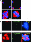
Simultaneous depletion of Rae1 and NuMA rescues bipolarity. (A) HeLa cells were transfected with either siRNA duplexes against Rae1 (Left) or Rae1 and NuMA together (Right). After 72 h, cells were stained with α-tubulin antibody (red) and analyzed by confocal laser microscopy. Chromatin was stained with DAPI (blue). [Scale bars, 25 μm (Upper); 5 μm (Lower).] (B and C) Representative figures of cells treated with Rae1 siRNA, fixed, and stained with anti-pericentrin and either γ-tubulin (B) or α-tubulin (C) antibodies. DNA is counterstained with DAPI.
Fig. 3.
Fig. 3.
Simultaneous overexpression of Rae1 and NuMA rescues bipolarity. Representative figures of HeLa cells transfected with plasmids overexpressing either GFP-NuMA or GFP-NuMA and Rae1-HA together. After 24 h, cells were fixed, stained with α-tubulin antibody (red in overlay; GFP is green), and analyzed by confocal laser microscopy. Chromatin was stained with DAPI (blue). (Scale bar, 5 μm.)
Fig. 4.
Fig. 4.
Mapping the Rae1 interaction domain on NuMA. (A) Schematic of NuMA and five FLAG-tagged fragments of NuMA. Numbers on the left refer to amino acids (aa); all fragments are continuous (e.g., NuMA2 ends at amino acid 829 and NuMA3 starts at amino acid 829). (B) Autoradiograph of [35S]methionine-labeled Rae1 and NuMA-FLAG fragments coexpressed in vitro, affinity-purified, and separated by SDS/PAGE. Rae1 is untagged. Asterisks indicate the five FLAG-tag NuMA fragments expressed in varying amounts using this system. Numbers indicate molecular weight markers in kilodaltons. (C) Immunoblotting of α-GFP IPs from either GFP- or GFP-NuMA325–829-expressing HeLa cells. IPs are blotted with α-Rae1 or α-NuMA [using BD Biosciences clone 22 monoclonal that recognizes an epitope (amino acids 658–691) within NuMA325–829]. (D) HeLa cells overexpressing GFP-NuMA325–829 (green) costained with tubulin (red) and DAPI (blue).
Fig. 5.
Fig. 5.
A “valency” model of MTs interacting with NuMA and Rae1. NuMA is assumed to be a dimer (6) with the C-terminal (C) indicated to directly interact with MTs (7). A region (residues 325–829) at the N-terminal end of the coiled coil of NuMA interacts with Rae1 (data in this paper) and therefore converts NuMA from a divalent to a tetravalent MT “crosslinker.”

References

    1. Compton DA. Annu Rev Biochem. 2000;69:95–114. - PubMed
    1. Merdes A, Cleveland DW. J Cell Biol. 1997;138:953–956. - PMC - PubMed
    1. Karsenti E, Vernos I. Science. 2001;294:543–547. - PubMed
    1. Wadsworth P, Khodjakov A. Trends Cell Biol. 2004;14:413–419. - PubMed
    1. Compton DA, Szilak I, Cleveland DW. J Cell Biol. 1992;116:1395–1408. - PMC - PubMed

Publication types

MeSH terms

Substances