. 2007 Feb;73(4):1180-8.
doi: 10.1128/AEM.01913-06. Epub 2006 Dec 15.

Identification of specific chemoattractants and genetic complementation of a Borrelia burgdorferi chemotaxis mutant: flow cytometry-based capillary tube chemotaxis assay

Affiliations

Identification of specific chemoattractants and genetic complementation of a Borrelia burgdorferi chemotaxis mutant: flow cytometry-based capillary tube chemotaxis assay

Richard G Bakker et al. Appl Environ Microbiol. 2007 Feb.

Abstract

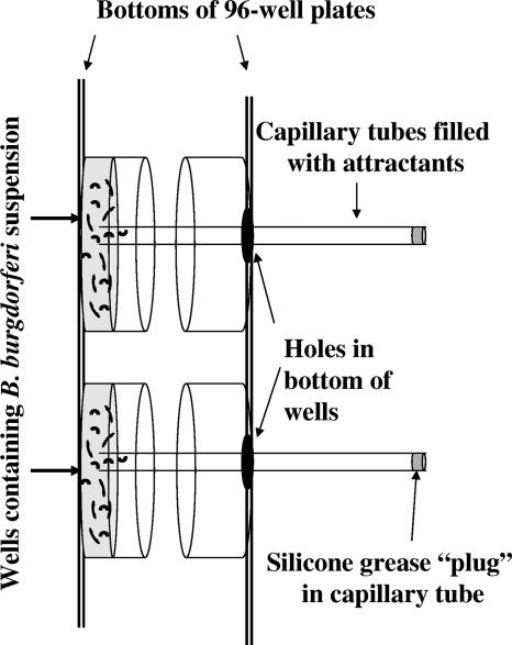
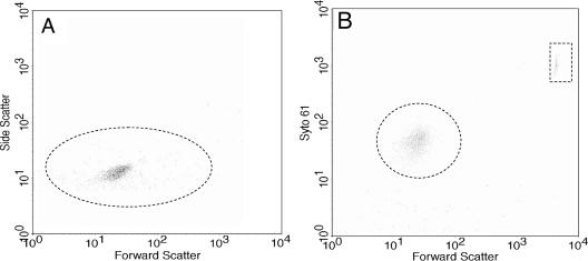
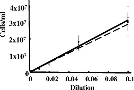
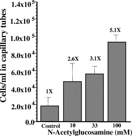
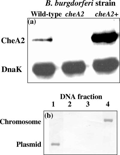
Measuring the chemotactic response of Borrelia burgdorferi, the bacterial species that causes Lyme disease, is relatively more difficult than measuring that of other bacteria. Because these spirochetes have long generation times, enumerating cells that swim up a capillary tube containing an attractant by using colony counts is impractical. Furthermore, direct counts with a Petroff-Hausser chamber is problematic, as this method has a low throughput and necessitates a high cell density; the latter can lead to misinterpretation of results when assaying for specific attractants. Only rabbit serum and tick saliva have been reported to be chemoattractants for B. burgdorferi. These complex biological mixtures are limited in their utility for studying chemotaxis on a molecular level. Here we present a modified capillary tube chemotaxis assay for B. burgdorferi that enumerates cells by flow cytometry. Initial studies identified N-acetylglucosamine as a chemoattractant. The assay was then optimized with respect to cell concentration, incubation time, motility buffer composition, and growth phase. Besides N-acetylglucosamine, glucosamine, glucosamine dimers (chitosan), glutamate, and glucose also elicited significant chemoattractant responses, although the response obtained with glucose was weak and variable. Serine and glycine were nonchemotactic. To further validate and to exploit the use of this assay, a previously described nonchemotactic cheA2 mutant was shown to be nonchemotactic by this assay; it also regained the wild-type phenotype when complemented in trans. This is the first report that identifies specific chemical attractants for B. burgdorferi and the use of flow cytometry for spirochete enumeration. The method should also be useful for assaying chemotaxis for other slow-growing prokaryotic species and in specific environments in nature.


Figures

FIG. 1.
FIG. 1.
Schematic of chemotaxis assay using two 96-well plates. B. burgdorferi cells in motility buffer plus methylcellulose were placed in wells of a 96-well plate. Holes were formed in the corresponding bottoms of wells of another 96-well plate with a heated metal cylinder. These 96-well plates were taped together face to face, with the plate containing the cells lying face up. Capillary tubes filled with attractant in motility buffer plus methylcellulose were plugged at one end with silicone grease and placed through the holes in the top 96-well plate. The ends of the capillary tubes dipped into the cell suspension in the corresponding wells of the other 96-well plate. The sandwiched plates were incubated such that the attractant-filled capillary tubes were horizontal but still remained inserted in the cell suspension.
FIG. 2.
FIG. 2.
Flow cytometric analysis of B. burgdorferi. Wild-type cells were analyzed by flow cytometry in the absence (A) or presence (B) of Syto61. The y axis represents side scatter in panel A and intensity of Syto61 staining in panel B. The dotted circle in panel A indicates that B. burgdorferi cells are detected by side scatter, and that in panel B indicates detection of cells stained with Syto61. Polystyrene beads formed a distinct fluorescent population (dotted box) in the presence of Syto61 (B). Results identical to those in panel B were also obtained with cheA2 mutant cells (not shown).
FIG. 3.
FIG. 3.
Comparison of cell enumeration by flow cytometry and the Petroff-Hausser counting chamber. B. burgdorferi bacteria were collected by centrifugation, suspended in motility buffer, and serially diluted. Cell concentrations were determined manually with a Petroff-Hausser counting chamber (squares, solid line) or by flow cytometric analysis (circles, dashed line). The coefficient of correlation between the two methods was >0.98.
FIG. 4.
FIG. 4.
Growth curve dependence of chemotaxis. B. burgdorferi cells were assayed for chemotaxis during different phases of growth. The chemotactic response is represented by the bars, and cell numbers per milliliter are represented by the dashed line. The cell division time during logarithmic growth was 9.33 h. Error lines represent SDs of quadruplicate samples.
FIG. 5.
FIG. 5.
Dependence of chemotaxis on N-acetylglucosamine concentration. Capillary tubes filled with motility buffer alone (control) or the indicated concentrations of N-acetylglucosamine were incubated with B. burgdorferi cells and assayed for chemotaxis. The results of a representative experiment are depicted, showing the mean and SD of quadruplicate samples. The relative chemotactic response is indicated as a times value at the top of each bar.
FIG. 6.
FIG. 6.
CheA2 and attractant gradient dependence of B. burgdorferi on chemotaxis to N-acetylglucosamine. Capillary tubes filled with motility buffer containing 100 mM N-acetylglucosamine were assayed for chemotaxis with wild-type B. burgdorferi, the cheA2 mutant, or complemented strain cheA2+. The relative chemotactic response compared to the buffer control was determined. Results are expressed as the mean of four experiments ± the standard error. “No gradient” indicates a control in which both the capillary tube and the suspending motility buffer contained equal concentrations of N-acetylglucosamine.
FIG. 7.
FIG. 7.
cheA2 inactivation and complementation. (a) Ten micrograms of protein from lysates of wild-type, cheA2, and cheA2+ cells were electrophoresed on sodium dodecyl sulfate-polyacrylamide gels, blotted onto nitrocellulose, and reacted with E. coli CheA and B. burgdorferi DnaK antibodies. (b) Southern blot analysis of chromosomal or plasmid DNA from cheA2+, wild-type, and cheA2 mutant cells (see the text). Southern blot analysis was carried out with a digoxigenin-labeled DNA probe complementary to a fragment of cheA2 deleted in the mutant (Table 1). The chromosomal and plasmid DNAs migrated at rates represented by the vertical axis.

References

    1. Adler, J. 1973. A method for measuring chemotaxis and use of the method to determine optimum conditions for chemotaxis by Escherichia coli. J. Gen. Microbiol. 74:77-91. - PubMed
    1. Babb, K., J. D. McAlister, J. C. Miller, and B. Stevenson. 2004. Molecular characterization of Borrelia burgdorferi erp promoter/operator elements. J. Bacteriol. 186:2745-2756. - PMC - PubMed
    1. Baker, M. D., P. M. Wolanin, and J. B. Stock. 2006. Signal transduction in bacterial chemotaxis. Bioessays 28:9-22. - PubMed
    1. Bakker, R. G. 2006. Measuring chemotaxis in Borrelia burgdorferi, the Lyme disease spirochete. Ph.D. dissertation. West Virginia University, Morgantown.
    1. Barbour, A. G. 1984. Isolation and cultivation of Lyme disease spirochetes. Yale J. Biol. Med. 57:521-525. - PMC - PubMed

Publication types