

doi: 10.1186/1747-1028-1-31.
Eg5 steps it up!
Affiliations
- PMID: 17173688
- PMCID: PMC1716758
- DOI: 10.1186/1747-1028-1-31
Item in Clipboard
Eg5 steps it up!
Cell Div.
.
Abstract
Understanding how molecular motors generate force and move microtubules in mitosis is essential to understanding the physical mechanism of cell division. Recent measurements have shown that one mitotic kinesin superfamily member, Eg5, is mechanically processive and capable of crosslinking and sliding microtubules in vitro. In this review, we highlight recent work that explores how Eg5 functions under load, with an emphasis on the nanomechanical properties of single enzymes.
Figures

Schematic depicting Eg5 activity in the mitotic spindle. Tetrameric Eg5 motors (red) help organize microtubules (green) to form the mitotic spindle. (A) At the onset of mitosis, the duplicated centrosomes (blue) separate and nucleate two microtubule asters. Processive Eg5 motors may translocate to the plus-ends of microtubules, located distal to the centrosomal organizing center and by crosslinking antiparallel microtubules, may promote bipolarity. (B) By metaphase, a stable bipolar spindle has formed. Eg5 motors likely provide structural integrity and also slide microtubules toward the centrosomes, contributing to the generation of poleward flux. (C) A close-up depiction of Eg5 motors walking to the plus ends of antiparallel microtubules, moving both poleward simultaneously.

Schematic showing in vitro assay designs for Eg5 motor studies. (A) Depiction of a fluorescence-based assay used to demonstrate purified full length Eg5 tetramers are capable of crosslinking and sliding microtubules in vitro [13]. Unlabeled Eg5 motors bind to fluorescent, polarity-marked microtubules, causing the microtubules to slide apart. (B) Schematic showing optical trapping assay used to observe processive movement of Eg5 dimers [30]. His-tagged motors are attached to streptavidin-coated beads through a biotinlyated PentaHis antibody. Coverslip surfaces are precoated with poly-L-lysine-graft-poly(ethylene glycol) polymers to prevent surface-induced denaturation of Eg5 at the glass interface. Polymers are biotinylated to allow the specific attachment of biotinlyated microtubules via a streptavidin linkage.
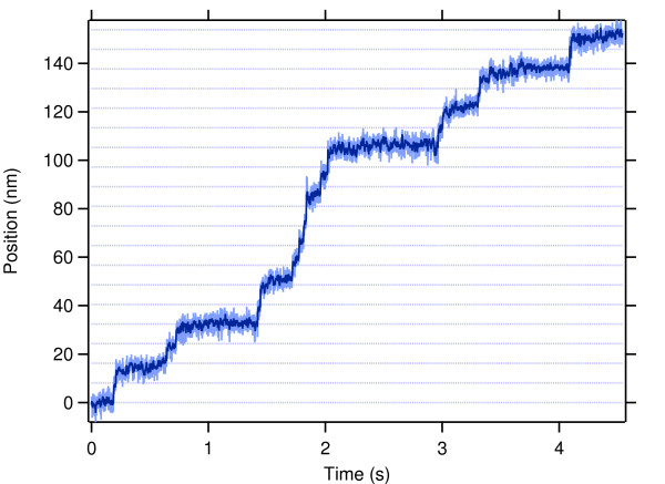
Representative trace of position of single Eg5 dimer moving in vitro. Record shows motion of a bead-attached Eg5 dimer held in an optical trap, and walking along microtubules in 8.1-nm steps. Position (light blue) and smoothed position (dark blue) are plotted as a function of time; dotted lines are placed every 8.1 nm to guide the eye. Experimental conditions: 2 mM ATP, 4 pN load applied toward the microtubule plus-end (assisting motion).

Comparison of the force-dependence of the velocities of Eg5 and conventional kinesin. Eg5 (red, left axis) [30] and conventional kinesin (blue, right axis) [37] velocity as a function of force, as measured with a force-clamped optical trap. Positive forces indicate that load was applied toward the plus-end of the microtubule, assisting motion; negative forces hinder translocation. Conventional kinesin slows much more dramatically than Eg5 does under hindering load.
References
Grants and funding
LinkOut - more resources
Full Text Sources
