

Comparative studies between species that do and do not exhibit the washout effect
- PMID: 17173894
- PMCID: PMC1850108
- DOI: 10.1016/j.exer.2006.10.015
Comparative studies between species that do and do not exhibit the washout effect
Abstract
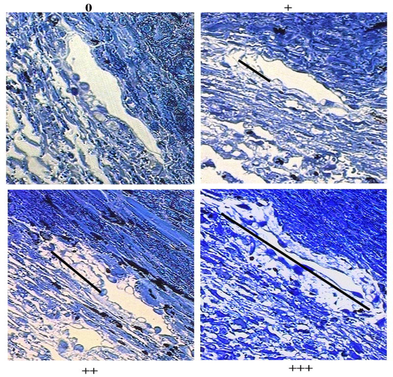
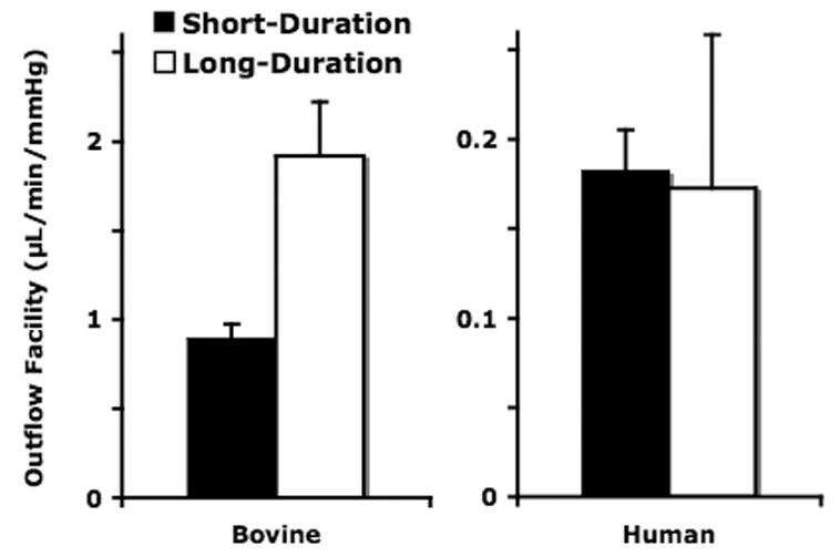
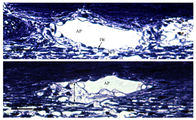
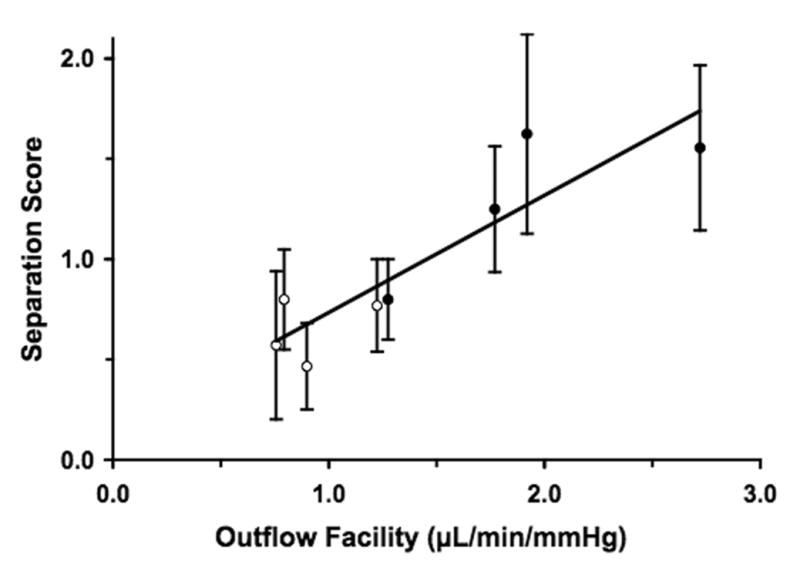
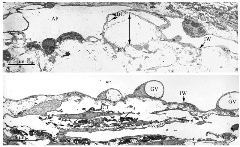
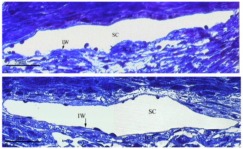
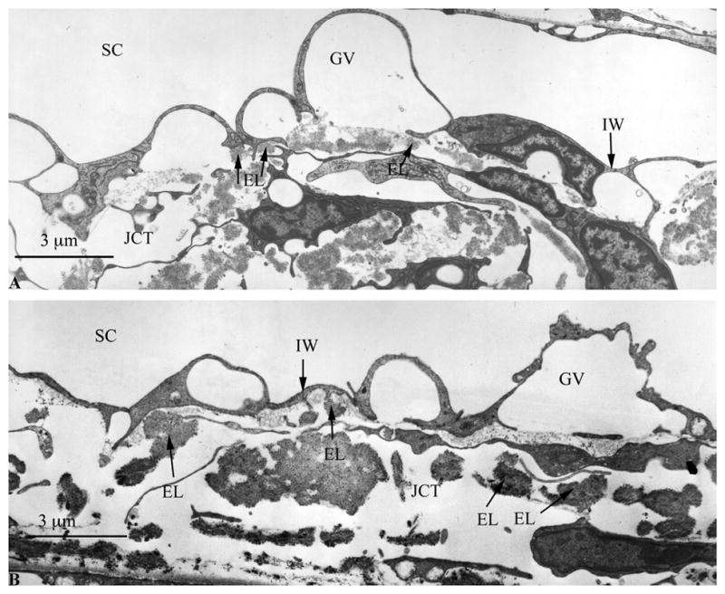
Ocular perfusion studies from all non-human species performed to date consistently demonstrate a perfusion-volume-dependent increase in aqueous outflow facility known as the "washout" effect. However, this "washout" effect does not occur in human eyes. We have recently documented that, in bovine eyes, the washout associated increase in facility correlates with the extent of physical separation between the juxtacanalicular connective tissue (JCT) and the inner wall endothelium lining the aqueous plexus (the bovine equivalent of Schlemm's canal). We hypothesize that if washout truly correlates with inner wall/JCT separation then this separation should not occur in human eyes that do not exhibit the washout effect, even after prolonged perfusion. Eight enucleated human and eight bovine eyes were used in this study. Aqueous humor outflow facility was measured at 15 mmHg for long-duration (3 h) or short-duration (30 min to 1 h) perfusion (n=4 for each group). All eyes were perfusion-fixed at 15 mmHg, and examined morphologically with both light and electron microscopy. In bovine eyes, outflow facility increased 81% (p=0.049) from 1.06 +/- 0.06 microl/min per mmHg (mean+/-SEM) at baseline to 1.92 +/- 0.30 microl/min per mmHg after 3 h due to washout. The pre-fixation outflow facility in long-duration eyes (1.92 +/- 0.30 microl/min per mmHg) was 2-fold greater than pre-fixation facility in short-duration eyes (0.92 +/- 0.11 microl/min per mmHg; p=0.0387). In human eyes, washout was not observed; baseline outflow facility was similar between both groups (0.18 +/- 0.02 vs. 0.25 +/- 0.08 microl/min per mmHg; p=0.518); however, pre-fixation outflow facility in long-duration eyes showed a 40% decrease compared to baseline outflow facility in those same eyes (p=0.017, paired Student's t-test). In bovine eyes, significant expansion and rarefaction of the JCT and inner wall/JCT separation was much more prevalent in long-duration eyes, and data from all bovine eyes revealed a correlation between the extent of inner wall/JCT separation and the absolute value of outflow facility measured immediately prior to fixation (p=0.0024) as well as the washout-induced increase in outflow facility (p=0.0006). In human eyes, no significant morphologic differences were observed between long- and short-duration perfusion, with no observed change in inner wall/JCT separation or expansion between the two groups. Morphologic analysis revealed that the previously described "cribriform plexus" of elastic-like fibers was far more extensive in the JCT of human eyes, appearing to form numerous connections to the inner wall endothelium. The cribriform plexus appears to function as a mechanical tether that maintains inner wall/JCT connectivity in human eyes by opposing hydrodynamic forces generated during perfusion, potentially explaining the lack of washout in humans.
Figures
Similar articles
-
Relationships between increased aqueous outflow facility during washout with the changes in hydrodynamic pattern and morphology in bovine aqueous outflow pathways.Exp Eye Res. 2009 Dec;89(6):942-9. doi: 10.1016/j.exer.2009.08.002. Epub 2009 Aug 11. Exp Eye Res. 2009. PMID: 19679123
-
Similar hydrodynamic and morphological changes in the aqueous humor outflow pathway after washout and Y27632 treatment in monkey eyes.Exp Eye Res. 2011 Oct;93(4):397-404. doi: 10.1016/j.exer.2011.05.012. Epub 2011 Jun 6. Exp Eye Res. 2011. PMID: 21669200 Free PMC article.
-
The mechanism of increasing outflow facility during washout in the bovine eye.Invest Ophthalmol Vis Sci. 2002 Nov;43(11):3455-64. Invest Ophthalmol Vis Sci. 2002. PMID: 12407156
-
The changing paradigm of outflow resistance generation: towards synergistic models of the JCT and inner wall endothelium.Exp Eye Res. 2009 Apr;88(4):656-70. doi: 10.1016/j.exer.2008.11.033. Epub 2008 Dec 11. Exp Eye Res. 2009. PMID: 19103197 Free PMC article. Review.
-
Morphological and hydrodynamic correlations with increasing outflow facility by rho-kinase inhibitor Y-27632.J Ocul Pharmacol Ther. 2014 Mar-Apr;30(2-3):143-53. doi: 10.1089/jop.2013.0192. Epub 2014 Jan 24. J Ocul Pharmacol Ther. 2014. PMID: 24460021 Free PMC article. Review.
Cited by
-
Functional Anatomy of the Outflow Facilities.Vet Clin North Am Small Anim Pract. 2015 Nov;45(6):1101-26, v. doi: 10.1016/j.cvsm.2015.06.005. Epub 2015 Aug 31. Vet Clin North Am Small Anim Pract. 2015. PMID: 26337760 Free PMC article. Review.
-
Preferential fluid flow in the human trabecular meshwork near collector channels.Invest Ophthalmol Vis Sci. 2009 Apr;50(4):1692-7. doi: 10.1167/iovs.08-2375. Epub 2008 Dec 5. Invest Ophthalmol Vis Sci. 2009. PMID: 19060275 Free PMC article.
-
Segmental Uveoscleral Outflow and its Relationship With Trabecular Outflow in Monkey Eyes.Invest Ophthalmol Vis Sci. 2025 Apr 1;66(4):78. doi: 10.1167/iovs.66.4.78. Invest Ophthalmol Vis Sci. 2025. PMID: 40293394 Free PMC article.
-
The Trabecular Meshwork: A Basic Review of Form and Function.J Ocul Biol. 2014 May;2(1):http://fulltextarticles.avensonline.org/JOCB-2334-2838-02-0017.html. doi: 10.13188/2334-2838.1000017. J Ocul Biol. 2014. PMID: 25356439 Free PMC article. No abstract available.
-
Dose- and time-dependent effects of actomyosin inhibition on live mouse outflow resistance and aqueous drainage tissues.Sci Rep. 2016 Feb 17;6:21492. doi: 10.1038/srep21492. Sci Rep. 2016. PMID: 26884319 Free PMC article.
References
-
- Bárány EH. In vitro studies of the resistance to flow through the angle of the anterior chamber. Acta Soc Med Uppsala. 1953;59:260–276. - PubMed
-
- Bárány EH. The mode of action of pilocarpine on outflow resistance in the eye of a primate (Cercopithecus aethiops) Invest Ophthalmol Vis Sci. 1962;1:712–727. - PubMed
-
- Bárány EH. Simultaneous measurement of changing intraocular pressure and outflow facility in the vervet monkey by constant pressure infusion. Invest Ophthalmol Vis Sci. 1964;3:135–143. - PubMed
-
- Bárány EH, Scotchbrook S. Influence of testicular hyaluronidase on the resistance to flow through the angle of the anterior chamber. Acta Physiol Scand. 1954;30:240–248. - PubMed
-
- Bárány EH, Woodin AM. Hyaluronic Acid and hyaluronidase in the aqueous humor and the angle of the anterior chamber. Acta Physiol Scand. 1955;33:257–290. - PubMed
Publication types
MeSH terms
Grants and funding
LinkOut - more resources
Full Text Sources
Other Literature Sources
