. 2007 Mar;84(3):444-54.
doi: 10.1016/j.exer.2006.10.020. Epub 2006 Dec 13.

4-Hydroxynonenal, a product of oxidative stress, leads to an antioxidant response in optic nerve head astrocytes

Affiliations

4-Hydroxynonenal, a product of oxidative stress, leads to an antioxidant response in optic nerve head astrocytes

P E Malone et al. Exp Eye Res. 2007 Mar.

Abstract

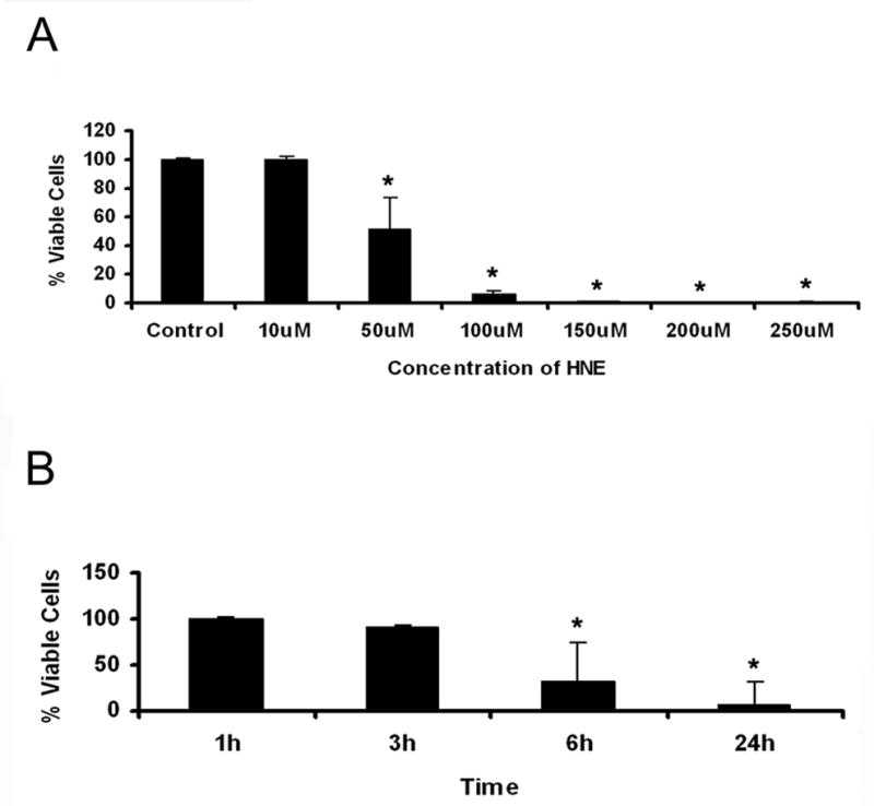
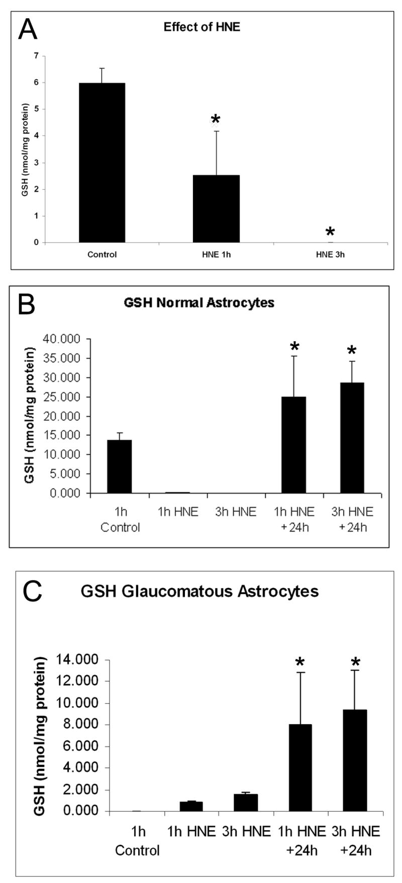
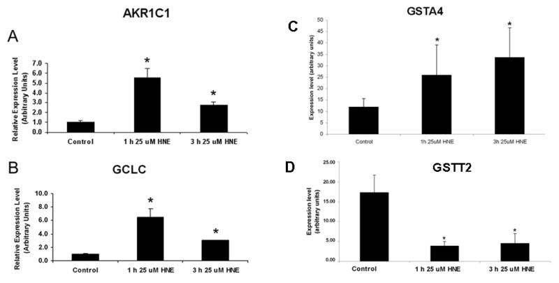
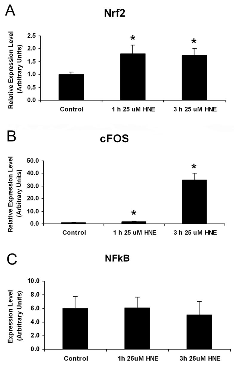
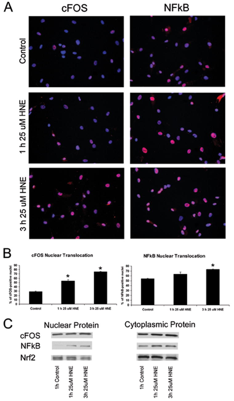
Oxidative stress has been implicated in the pathogenesis of several neurodegenerative disorders including primary open-angle glaucoma (POAG) an optic neuropathy characterized by loss of retinal ganglion cell (RGC) axons and remodeling of the optic nerve head (ONH). Previous findings in glaucomatous astrocytes suggested increased oxidative stress and lipid peroxidation in human optic nerves. We studied the dose and time dependent effects of 4-hydroxynonenal (HNE), a by-product of lipid peroxidation, on the viability of primary cultures of human ONH astrocyte. A significant depletion of glutathione (GSH) level was observed in normal astrocytes after exposure to HNE for 1 h and 3 h. Untreated glaucomatous astrocytes exhibited depleted levels of GSH which increased slightly after exposure to HNE. Both normal and glaucomatous astrocytes recovered GSH levels after 24 h of removal of HNE. HNE caused significant increases in expression of antioxidant enzymes, glutamate cysteine ligase catalytic subunit (GCLC), aldo-keto reductase 1C family member 1 (AKR1C1) and glutathione S-transferase-alpha4 (GSTA4). HNE induced expression of the transcription factor Nrf2, which coordinates the upregulation of detoxification enzymes. In addition, ONH astrocytes responded to HNE by activation and transcription of cFOS and NFkB, which regulate physiological protective responses against oxidative stress. Our results indicate that ONH astrocytes exhibit a strong antioxidant response to HNE treatment by inducing the transcription factors cFOS, NFkB, and Nrf2, which upregulate the expression of GCLC, to produce more GSH in the cell. AKR1C1 was also upregulated after HNE treatment to inactivate HNE, independent of GSH availability in the cells. Collectively these data indicate that ONH astrocytes can efficiently counteract the neurotoxic effects of HNE offering protection in the optic nerve by releasing GSH and antioxidant enzymes to eliminate the products of chronic oxidative stress.


Figures

Figure 1
Figure 1. A. Cell viability assay
Normal ONH astrocytes were treated with varying concentrations of HNE for 6 h. Treatment with HNE at concentrations of 50 μM and higher led to a greater than 50% reduction in cell viability over 6 h. The data is expressed as % of viable cells of the untreated control. Asterisks indicate significant difference (*P<0.05). B. Time course of HNE (25 μM) treatment. Normal ONH astrocytes were treated with 25 μM HNE for different exposure times. Treatment with 25 μM HNE for 3 h led to a 10% reduction in cell viability and was used in future experiments. Asterisks indicate significant difference (*P<0.05).
Figure 2
Figure 2. A. Effect of HNE on Glutathione (GSH) content of normal astrocytes
Normal ONH astrocytes from a 29 year old donor were treated with 25 μM HNE for 1 h and 3 H. In normal ONH astrocytes levels of GSH become depleted after 25 μM HNE treatment. Asterisks indicate significant difference (*P<0.05). B. Glutathione (GSH) content of normal astrocytes. Normal ONH astrocytes from a 65 year old donor were treated with 25 μM HNE for 1 h and 3 h, and GSH content was measured immediately after treatment, as well as after a 24 h recovery without HNE-containing medium following each treatment duration. Levels of GSH become depleted after 25 μM HNE treatment for 1h and 3h in normal astrocytes. After a 24 h recovery from HNE treatment, the levels of GSH are over 2-fold higher than basal levels. Asterisks indicate significant difference (*P<0.05) from control and from treatment with HNE. C. Glutathione (GSH) content of glaucomatous astrocytes. Glaucomatous ONH astrocytes from a 70 year old donor were treated with 25 μM HNE for 1 h and 3 h, and GSH content was measured immediately after treatment, as well as after a 24 h recovery without HNE-containing medium following each treatment duration. Glaucomatous astrocytes exhibit basal levels of GSH below the level of detection compared to normal astrocytes, which increase slightly after 25 μM HNE treatment for 1h and 3h. After a 24 h recovery from HNE treatment, the levels of GSH are higher. Asterisks indicate significant difference (*P<0.05) from control and from treatment with HNE.
Figure 3
Figure 3. Effects of exposure to HNE in gene expression of antioxidant enzymes
Normal astrocytes were treated for 1 h and 3 h with 25 μM HNE, then the HNE-containing media was removed and RNA was collected 6 h later. Relative amount of target mRNA in ONH astrocytes measured by quantitative RT-PCR as described in the Methods. Values represent mean ±SD of target mRNA level normalized to 18S RNA (internal control). All experiments were done in triplicate using three normal astrocytes cultures. Asterisk indicates significance of P <0.05 A. Expression of AKR1C1. The relative expression of AKR1C1 mRNA is 5.56 times higher after 1 h HNE treatment, and 2.77 times higher after 3 h HNE treatment, when compared to controls. B. Expression of GCLC. The relative expression of GCLC mRNA increased after 1 h HNE treatment to 6.49 times higher than control levels, and to a lesser extent after 3 h HNE treatment to 3.02 times higher than control levels. C. Expression of GSTA4. The relative expression of GSTA4 mRNA increased after 1 h HNE treatment to 2.2 times higher than control levels, and 2.8 times higher than control levels after 3 h HNE treatment. D. Expression of GSTT2. The relative expression of GSTT2 mRNA was 4.60 times lower than control levels after 1 h HNE treatment to, and 3.89 times lower than control levels after 3 h HNE treatment.
Figure 4
Figure 4. Effects of HNE on transcription factor gene expression
Normal astrocytes were treated for 1 h and 3 h with 25 μM HNE, then the HNE-containing media was removed and RNA was collected 6 h later. Relative amount of target mRNA in ONH astrocytes measured by quantitative RT-PCR as described in the Methods. Values represent mean ±SD of target mRNA level normalized to 18S RNA (internal control). All experiments were done in triplicate using three normal astrocytes cultures. Asterisk indicates significance of P <0.05. A. Increased expression of Nrf2 using qRT-PCR. The relative expression of Nrf2 mRNA increased in response to both a 1 h and 3 h HNE treatment to 1.80 and 1.75 times higher than control levels, respectively. B. Induction of cFOS expression. The relative expression of cFOS mRNA increased after 1 h and 3 h HNE. A smaller increase of cFOS mRNA occurred after 1 h HNE treatment to 1.86 times higher than control levels, and a great increase of cFOS mRNA to 34.96 times higher than control levels occurred after 3 h HNE treatment. C. Expression of NFkB. The relative expression of NFkB did not change after treatment with HNE. All experiments were done in triplicate using three normal astrocytes cultures.
Figure 5
Figure 5. HNE affects the nuclear localization of c-Fos in normal human ONH astrocytes
A. cFOS and NFkB nuclear localization. Treatment of normal ONH astrocytes with 25 μM HNE for 1 h and 3 h led to nuclear localization of both cFOS and NFkB, identified by immunofluorescence staining. cFOS and NFkB are stained in red; nuclei are stained in blue with DAPI, and localization of cFOS or NFkB to the nucleus results in a pink staining of nuclei. B. cFOS and NFkB nuclear translocation measurements. Treatment of normal ONH astrocytes with 25 μM HNE led to nuclear translocation of cFOS and NFkB. Percentage of nuclear localization of cFOS in control astrocytes was 28.82%. Upon exposure to HNE for 1h and 3 h, c-Fos nuclear localization increased to 53.67%, and 75.07%, respectively. Percent of NFkB-positive nuclei in control, 1 h HNE, and 3 h HNE-treated astrocytes were 54.15%, 63.46%, and 73.29%, respectively. Asterisk indicates significance of P <0.05. All experiments were done in triplicate I ONH astrocytes fro three normal donors. C. Western blots of nuclear and cytoplasmic protein. Normal astrocytes were treated for 1 h and 3 h with 25 μM HNE, then the HNE-containing media was removed and protein was collected 6 h later. In controls we detected abundant cFOS protein in the nucleus and cytoplasm. No discernable changes in cFOS cytoplasmic or nuclear protein were detected, nor any changes in NFkB cytoplasmic protein levels. In controls there were very low levels of NFkB protein detected in the nucleus which increased after treatment for 1 h and 3 h with 25 μM HNE. Nrf2 protein was detected in the nucleus and cytoplasm but there were no changes after exposure to HNE.

References

    1. Agapova OA, Ricard CS, Salvador-Silva M, Hernandez MR. Expression of matrix metalloproteinases and tissue inhibitors of metalloproteinases in human optic nerve head astrocytes. Glia. 2001;33:205–216. - PubMed
    1. Agapova OA, Yang P, Wang WH, Lane DA, Clark AF, Weinstein BI, Hernandez MR. Altered expression of 3 alpha-hydroxysteroid dehydrogenases in human glaucomatous optic nerve head astrocytes. Neurobiol Dis. 2003;14:63–73. - PubMed
    1. Ahmed I, John A, Vijayasarathy C, Robin MA, Raza H. Differential modulation of growth and glutathione metabolism in cultured rat astrocytes by 4-hydroxynonenal and green tea polyphenol, epigallocatechin-3-gallate. Neurotoxicology. 2002;23:289–300. - PubMed
    1. Bolanos JP, Heales SJ, Peuchen S, Barker JE, Land JM, Clark JB. Nitric oxide-mediated mitochondrial damage: a potential neuroprotective role for glutathione. Free Radic Biol Med. 1996;21:995–1001. - PubMed
    1. Burczynski ME, Sridhar GR, Palackal NT, Penning TM. The reactive oxygen species--and Michael acceptor-inducible human aldoketo reductase AKR1C1 reduces the alpha,beta-unsaturated aldehyde 4-hydroxy-2-nonenal to 1,4-dihydroxy-2-nonene. J Biol Chem. 2001;276:2890–2897. - PubMed

Publication types

MeSH terms